{"title":"Spinopelvic sagittal realignment and incidence of adjacent segment disease after single-segment posterior lumbar inter-body fusion using 12° lordotic cages-a 2-year prospective cohort study.","authors":"Tomiya Matsumoto, Shinya Okuda, Yukitaka Nagamoto, Yoshifumi Takahashi, Masayuki Furuya, Motoki Iwasaki","doi":"10.21037/jss-23-78","DOIUrl":null,"url":null,"abstract":"<p><strong>Background: </strong>The importance of spinopelvic sagittal alignment for adjacent segment disease (ASD) after lumbar fusion surgery has been reported. However, no longitudinal cohort studies have determined the extent to which segmental alignment and spinopelvic global alignment can be achieved using 12° lordotic cages in posterior lumbar inter-body fusion (PLIF) and the extent to which the development of ASD can be prevented. The purpose of this study was to analyze changes in segmental and spinopelvic sagittal alignment after single-segment PLIF with 12° lordotic cages, to clarify the relationship between changes in segmental and spinopelvic sagittal alignment, and to report the incidence of ASD at 2 years postoperatively.</p><p><strong>Methods: </strong>Subjects in this 2-year prospective longitudinal cohort study were 28 patients who had undergone L4/5 PLIF using 12° lordotic cages. Incidence of operative ASD (O-ASD) was evaluated as clinical outcomes. Radiological measurements were examined preoperatively and at 3 months, 1 year and 2 years postoperatively. The following radiographic spinopelvic parameters were measured: segmental lordosis (SL) at L4/5; sagittal vertical axis (SVA); T1 pelvic angle (TPA); thoracic kyphosis (TK); lumbar lordosis (LL); sacral slope (SS); pelvic tilt (PT); and pelvic incidence (PI). With respect to radiological outcomes, changes in SL (ΔSL) and spinopelvic parameters and the incidence of radiological ASD (R-ASD) were evaluated. Correlations of ΔSL and changes in other spinopelvic parameters (ΔSVA, ΔTPA, ΔTK, ΔLL, ΔSS, ΔPT, and ΔPI-LL) between preoperatively and 3 months postoperatively were examined.</p><p><strong>Results: </strong>The follow-up rate was 100% (n=28) at 1 year postoperatively and 96.4% (n=27) at 2 years postoperatively. No cases of O-ASD were seen during 2 years of follow-up. Significant realignment was observed and maintained at 2 years postoperatively in almost all spinopelvic sagittal parameters (SL, SVA, TPA, LL, PT, PI-LL). Regarding the correlation between ΔSL and other parameters, significant correlations were detected with ΔSVA (r=-0.37, P<0.05) and ΔLL (r=0.538, P<0.01). Three cases (11.1%) showed R-ASD at 2 years postoperatively.</p><p><strong>Conclusions: </strong>PLIF with 12° lordotic cages for L4 degenerative spondylolisthesis improved SL and global sagittal realignment, and achieved satisfactory clinical outcomes with a low incidence of ASD during 2 years of follow-up.</p>","PeriodicalId":17131,"journal":{"name":"Journal of spine surgery","volume":"9 3","pages":"269-277"},"PeriodicalIF":0.0000,"publicationDate":"2023-09-22","publicationTypes":"Journal Article","fieldsOfStudy":null,"isOpenAccess":false,"openAccessPdf":"https://ftp.ncbi.nlm.nih.gov/pub/pmc/oa_pdf/69/c1/jss-09-03-269.PMC10570649.pdf","citationCount":"0","resultStr":null,"platform":"Semanticscholar","paperid":null,"PeriodicalName":"Journal of spine surgery","FirstCategoryId":"1085","ListUrlMain":"https://doi.org/10.21037/jss-23-78","RegionNum":0,"RegionCategory":null,"ArticlePicture":[],"TitleCN":null,"AbstractTextCN":null,"PMCID":null,"EPubDate":"2023/9/18 0:00:00","PubModel":"Epub","JCR":"Q1","JCRName":"Medicine","Score":null,"Total":0}
引用次数: 0
Abstract
Background: The importance of spinopelvic sagittal alignment for adjacent segment disease (ASD) after lumbar fusion surgery has been reported. However, no longitudinal cohort studies have determined the extent to which segmental alignment and spinopelvic global alignment can be achieved using 12° lordotic cages in posterior lumbar inter-body fusion (PLIF) and the extent to which the development of ASD can be prevented. The purpose of this study was to analyze changes in segmental and spinopelvic sagittal alignment after single-segment PLIF with 12° lordotic cages, to clarify the relationship between changes in segmental and spinopelvic sagittal alignment, and to report the incidence of ASD at 2 years postoperatively.
Methods: Subjects in this 2-year prospective longitudinal cohort study were 28 patients who had undergone L4/5 PLIF using 12° lordotic cages. Incidence of operative ASD (O-ASD) was evaluated as clinical outcomes. Radiological measurements were examined preoperatively and at 3 months, 1 year and 2 years postoperatively. The following radiographic spinopelvic parameters were measured: segmental lordosis (SL) at L4/5; sagittal vertical axis (SVA); T1 pelvic angle (TPA); thoracic kyphosis (TK); lumbar lordosis (LL); sacral slope (SS); pelvic tilt (PT); and pelvic incidence (PI). With respect to radiological outcomes, changes in SL (ΔSL) and spinopelvic parameters and the incidence of radiological ASD (R-ASD) were evaluated. Correlations of ΔSL and changes in other spinopelvic parameters (ΔSVA, ΔTPA, ΔTK, ΔLL, ΔSS, ΔPT, and ΔPI-LL) between preoperatively and 3 months postoperatively were examined.
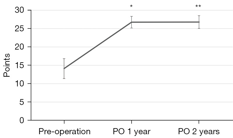
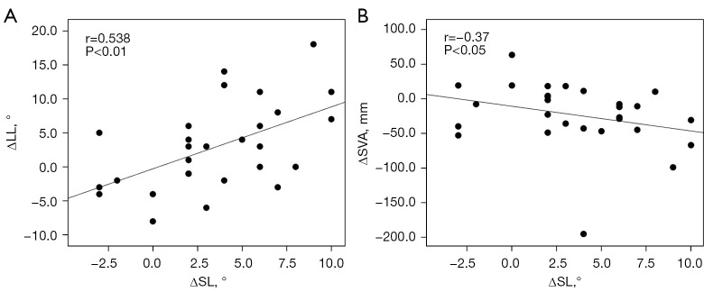
Results: The follow-up rate was 100% (n=28) at 1 year postoperatively and 96.4% (n=27) at 2 years postoperatively. No cases of O-ASD were seen during 2 years of follow-up. Significant realignment was observed and maintained at 2 years postoperatively in almost all spinopelvic sagittal parameters (SL, SVA, TPA, LL, PT, PI-LL). Regarding the correlation between ΔSL and other parameters, significant correlations were detected with ΔSVA (r=-0.37, P<0.05) and ΔLL (r=0.538, P<0.01). Three cases (11.1%) showed R-ASD at 2 years postoperatively.
Conclusions: PLIF with 12° lordotic cages for L4 degenerative spondylolisthesis improved SL and global sagittal realignment, and achieved satisfactory clinical outcomes with a low incidence of ASD during 2 years of follow-up.

 求助内容:
求助内容: 应助结果提醒方式:


