Jung-Hwa Tao-Cheng, Sandra L Moreira, Christine A Winters, Thomas S Reese, Ayse Dosemeci
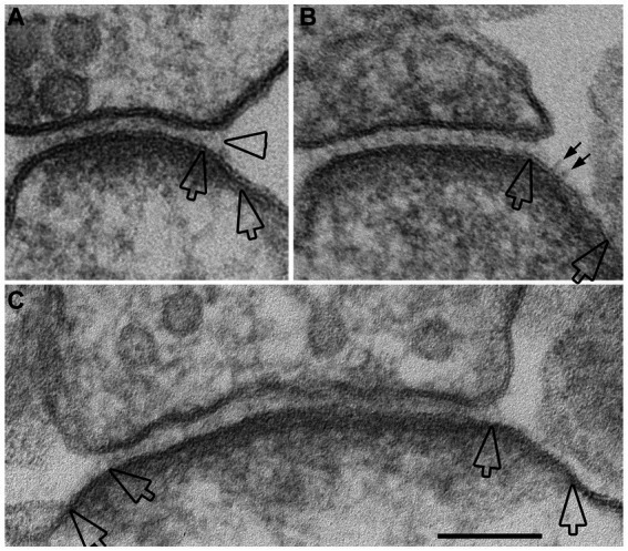
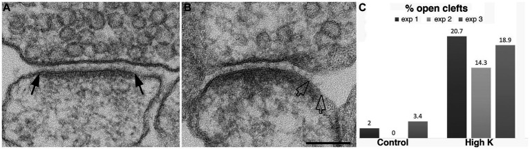
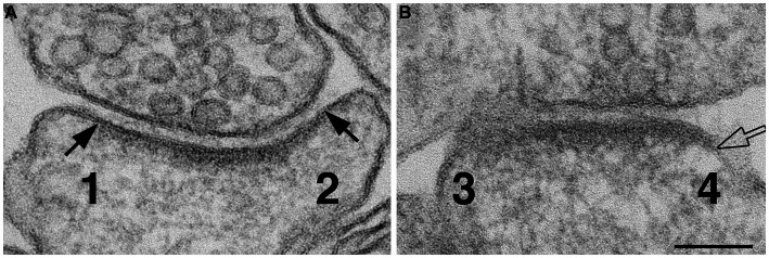
{"title":"Modification of the synaptic cleft under excitatory conditions.","authors":"Jung-Hwa Tao-Cheng, Sandra L Moreira, Christine A Winters, Thomas S Reese, Ayse Dosemeci","doi":"10.3389/fnsyn.2023.1239098","DOIUrl":null,"url":null,"abstract":"<p><p>The synaptic cleft is the extracellular part of the synapse, bridging the pre- and postsynaptic membranes. The geometry and molecular organization of the cleft is gaining increased attention as an important determinant of synaptic efficacy. The present study by electron microscopy focuses on short-term morphological changes at the synaptic cleft under excitatory conditions. Depolarization of cultured hippocampal neurons with high K<sup>+</sup> results in an increased frequency of synaptic profiles with clefts widened at the periphery (open clefts), typically exhibiting patches of membranes lined by postsynaptic density, but lacking associated presynaptic membranes (18.0% open clefts in high K<sup>+</sup> compared to 1.8% in controls). Similarly, higher frequencies of open clefts were observed in adult brain upon a delay of perfusion fixation to promote excitatory/ischemic conditions. Inhibition of basal activity in cultured neurons through the application of TTX results in the disappearance of open clefts whereas application of NMDA increases their frequency (19.0% in NMDA vs. 5.3% in control and 2.6% in APV). Depletion of extracellular Ca<sup>2+</sup> with EGTA also promotes an increase in the frequency of open clefts (16.6% in EGTA vs. 4.0% in controls), comparable to that by depolarization or NMDA, implicating dissociation of Ca<sup>2+</sup>-dependent trans-synaptic bridges. Dissociation of transsynaptic bridges under excitatory conditions may allow perisynaptic mobile elements, such as AMPA receptors to enter the cleft. In addition, peripheral opening of the cleft would facilitate neurotransmitter clearance and thus may have a homeostatic and/or protective function.</p>","PeriodicalId":12650,"journal":{"name":"Frontiers in Synaptic Neuroscience","volume":"15 ","pages":"1239098"},"PeriodicalIF":4.1000,"publicationDate":"2023-09-28","publicationTypes":"Journal Article","fieldsOfStudy":null,"isOpenAccess":false,"openAccessPdf":"https://www.ncbi.nlm.nih.gov/pmc/articles/PMC10568020/pdf/","citationCount":"0","resultStr":null,"platform":"Semanticscholar","paperid":null,"PeriodicalName":"Frontiers in Synaptic Neuroscience","FirstCategoryId":"3","ListUrlMain":"https://doi.org/10.3389/fnsyn.2023.1239098","RegionNum":4,"RegionCategory":"医学","ArticlePicture":[],"TitleCN":null,"AbstractTextCN":null,"PMCID":null,"EPubDate":"2023/1/1 0:00:00","PubModel":"eCollection","JCR":"Q2","JCRName":"NEUROSCIENCES","Score":null,"Total":0}
引用次数: 0
Abstract
The synaptic cleft is the extracellular part of the synapse, bridging the pre- and postsynaptic membranes. The geometry and molecular organization of the cleft is gaining increased attention as an important determinant of synaptic efficacy. The present study by electron microscopy focuses on short-term morphological changes at the synaptic cleft under excitatory conditions. Depolarization of cultured hippocampal neurons with high K+ results in an increased frequency of synaptic profiles with clefts widened at the periphery (open clefts), typically exhibiting patches of membranes lined by postsynaptic density, but lacking associated presynaptic membranes (18.0% open clefts in high K+ compared to 1.8% in controls). Similarly, higher frequencies of open clefts were observed in adult brain upon a delay of perfusion fixation to promote excitatory/ischemic conditions. Inhibition of basal activity in cultured neurons through the application of TTX results in the disappearance of open clefts whereas application of NMDA increases their frequency (19.0% in NMDA vs. 5.3% in control and 2.6% in APV). Depletion of extracellular Ca2+ with EGTA also promotes an increase in the frequency of open clefts (16.6% in EGTA vs. 4.0% in controls), comparable to that by depolarization or NMDA, implicating dissociation of Ca2+-dependent trans-synaptic bridges. Dissociation of transsynaptic bridges under excitatory conditions may allow perisynaptic mobile elements, such as AMPA receptors to enter the cleft. In addition, peripheral opening of the cleft would facilitate neurotransmitter clearance and thus may have a homeostatic and/or protective function.

 求助内容:
求助内容: 应助结果提醒方式:


