A reproducibility study of knee cartilage volume and thickness values derived by fully automatic segmentation based on three-dimensional dual-echo in steady state data from 1.5 T and 3 T magnetic resonance imaging.
IF 2 4区 医学Q3 RADIOLOGY, NUCLEAR MEDICINE & MEDICAL IMAGING
{"title":"A reproducibility study of knee cartilage volume and thickness values derived by fully automatic segmentation based on three-dimensional dual-echo in steady state data from 1.5 T and 3 T magnetic resonance imaging.","authors":"Ranxu Zhang, Xiaoyue Zhou, Esther Raithel, Congcong Ren, Ping Zhang, Junfei Li, Lin Bai, Jian Zhao","doi":"10.1007/s10334-023-01122-x","DOIUrl":null,"url":null,"abstract":"<p><strong>Objective: </strong>To evaluate the repeatability of cartilage volume and thickness values at 1.5 T MRI using a fully automatic cartilage segmentation method and reproducibility of the method between 1.5 T and 3 T data.</p><p><strong>Methods: </strong>The study included 20 knee joints from 10 healthy subjects with each subject having undergone double-knee MRI. All knees were scanned at 1.5 T and 3 T MR scanners using a three-dimensional (3D) high-resolution dual-echo in steady state (DESS) sequence. Cartilage volume and thickness of 21 subregions were quantified using a fully automatic cartilage segmentation research application (MR Chondral Health, version 3.0, Siemens Healthcare, Erlangen, Germany). The volume and thickness values derived from fully automatically computed segmentation masks were analyzed for the scan-rescan data from the same volunteers. The accuracy of the automatic segmentation of the cartilage in 1.5 T images was evaluated by the dice similarity coefficient (DSC) and Hausdorff distance (HD) using the manually corrected segmentation as a reference. The volume and thickness values calculated from 1.5 T and 3 T were also compared.</p><p><strong>Results: </strong>No statistically significant differences were found for cartilage thickness or volume across all subregions between the scan-rescanned data at 1.5 T (P > 0.05). The mean DSC between the fully automatic and manually corrected knee cartilage segmentation contours at 1.5 T was 0.9946. The average value of HD was 2.41 mm. Overall, there was no statistically significant difference in the cartilage volume or thickness in most-subregions between the two field strengths (P > 0.05) except for the medial region of femur and tibia. Bland-Altman plot and intraclass correlation coefficient (ICC) showed high consistency between results obtained based on same and different scanning sequences.</p><p><strong>Conclusion: </strong>The cartilage segmentation software had high repeatability for DESS images obtained from the same device. In addition, the overall reproducibility of the images obtained from equipment of two different field strengths was satisfactory.</p>","PeriodicalId":18067,"journal":{"name":"Magnetic Resonance Materials in Physics, Biology and Medicine","volume":" ","pages":"69-82"},"PeriodicalIF":2.0000,"publicationDate":"2024-02-01","publicationTypes":"Journal Article","fieldsOfStudy":null,"isOpenAccess":false,"openAccessPdf":"","citationCount":"0","resultStr":null,"platform":"Semanticscholar","paperid":null,"PeriodicalName":"Magnetic Resonance Materials in Physics, Biology and Medicine","FirstCategoryId":"3","ListUrlMain":"https://doi.org/10.1007/s10334-023-01122-x","RegionNum":4,"RegionCategory":"医学","ArticlePicture":[],"TitleCN":null,"AbstractTextCN":null,"PMCID":null,"EPubDate":"2023/10/10 0:00:00","PubModel":"Epub","JCR":"Q3","JCRName":"RADIOLOGY, NUCLEAR MEDICINE & MEDICAL IMAGING","Score":null,"Total":0}
引用次数: 0
Abstract
Objective: To evaluate the repeatability of cartilage volume and thickness values at 1.5 T MRI using a fully automatic cartilage segmentation method and reproducibility of the method between 1.5 T and 3 T data.
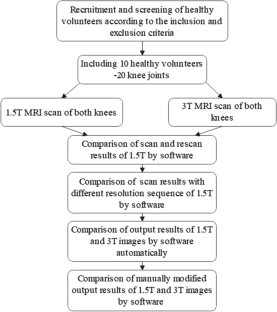
Methods: The study included 20 knee joints from 10 healthy subjects with each subject having undergone double-knee MRI. All knees were scanned at 1.5 T and 3 T MR scanners using a three-dimensional (3D) high-resolution dual-echo in steady state (DESS) sequence. Cartilage volume and thickness of 21 subregions were quantified using a fully automatic cartilage segmentation research application (MR Chondral Health, version 3.0, Siemens Healthcare, Erlangen, Germany). The volume and thickness values derived from fully automatically computed segmentation masks were analyzed for the scan-rescan data from the same volunteers. The accuracy of the automatic segmentation of the cartilage in 1.5 T images was evaluated by the dice similarity coefficient (DSC) and Hausdorff distance (HD) using the manually corrected segmentation as a reference. The volume and thickness values calculated from 1.5 T and 3 T were also compared.
Results: No statistically significant differences were found for cartilage thickness or volume across all subregions between the scan-rescanned data at 1.5 T (P > 0.05). The mean DSC between the fully automatic and manually corrected knee cartilage segmentation contours at 1.5 T was 0.9946. The average value of HD was 2.41 mm. Overall, there was no statistically significant difference in the cartilage volume or thickness in most-subregions between the two field strengths (P > 0.05) except for the medial region of femur and tibia. Bland-Altman plot and intraclass correlation coefficient (ICC) showed high consistency between results obtained based on same and different scanning sequences.
Conclusion: The cartilage segmentation software had high repeatability for DESS images obtained from the same device. In addition, the overall reproducibility of the images obtained from equipment of two different field strengths was satisfactory.
期刊介绍:
MAGMA is a multidisciplinary international journal devoted to the publication of articles on all aspects of magnetic resonance techniques and their applications in medicine and biology. MAGMA currently publishes research papers, reviews, letters to the editor, and commentaries, six times a year. The subject areas covered by MAGMA include:
advances in materials, hardware and software in magnetic resonance technology,
new developments and results in research and practical applications of magnetic resonance imaging and spectroscopy related to biology and medicine,
study of animal models and intact cells using magnetic resonance,
reports of clinical trials on humans and clinical validation of magnetic resonance protocols.

 求助内容:
求助内容: 应助结果提醒方式:


