Michael C Granovetter, Anne Margarette S Maallo, Christina Patterson, Daniel Glen, Marlene Behrmann
{"title":"Morphometrics of the preserved post-surgical hemisphere in pediatric drug-resistant epilepsy and implications for post-operative cognition.","authors":"Michael C Granovetter, Anne Margarette S Maallo, Christina Patterson, Daniel Glen, Marlene Behrmann","doi":"10.1101/2023.09.24.559189","DOIUrl":null,"url":null,"abstract":"<p><p>Characterization of the structural integrity of cortex in adults who have undergone resection for epilepsy treatment has, in some cases, revealed persistent or even accelerated cortical atrophy but, in others, the converse is evident, and atrophy decelerates or even reverses. Whether this variability applies to a pediatric population, for whom postoperative plasticity may be greater than in adulthood, remains to be determined. Furthermore, understanding the morphometrics of this patient population is important, as cognitive gains have been associated with the anatomical status of preserved cortex post-resection. Here, we used high-resolution structural T1 magnetic resonance imaging data to compare the (1) gross anatomy, (2) cortical thickness, volume, and surface area for 34 cortical regions, and (3) volume for nine subcortical regions of 32 pediatric post-surgical cases and 51 healthy controls. Patients with either a preserved right hemisphere (RH) or left hemisphere (LH) had lower total white matter volume and select subcortical structures' volumes, relative to controls; lateral ventricle size of both preserved RH and LH patients was also significantly larger than that of controls. However, relative to controls, only patients with a preserved RH had significantly lower total gray matter volume and lower thickness, volume, and surface area in multiple cortical regions, primarily in frontal and temporal cortex. The differences in preserved RH cortex of LH resection patients may relate to transfer of language function from the resected LH. Our findings lay the foundation for future studies probing associations of the morphometric differences in pediatric epilepsy surgery patients with neuropsychological outcomes.</p>","PeriodicalId":72407,"journal":{"name":"bioRxiv : the preprint server for biology","volume":" ","pages":""},"PeriodicalIF":0.0000,"publicationDate":"2025-08-25","publicationTypes":"Journal Article","fieldsOfStudy":null,"isOpenAccess":false,"openAccessPdf":"https://www.ncbi.nlm.nih.gov/pmc/articles/PMC10557613/pdf/","citationCount":"0","resultStr":null,"platform":"Semanticscholar","paperid":null,"PeriodicalName":"bioRxiv : the preprint server for biology","FirstCategoryId":"1085","ListUrlMain":"https://doi.org/10.1101/2023.09.24.559189","RegionNum":0,"RegionCategory":null,"ArticlePicture":[],"TitleCN":null,"AbstractTextCN":null,"PMCID":null,"EPubDate":"","PubModel":"","JCR":"","JCRName":"","Score":null,"Total":0}
引用次数: 0
Abstract
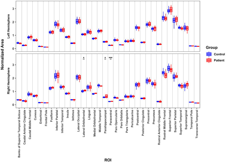
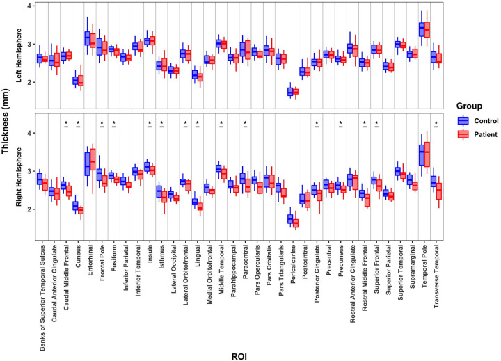
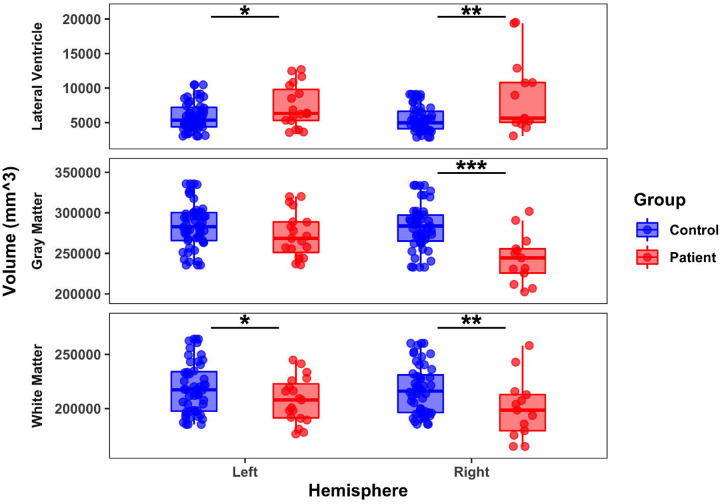
Characterization of the structural integrity of cortex in adults who have undergone resection for epilepsy treatment has, in some cases, revealed persistent or even accelerated cortical atrophy but, in others, the converse is evident, and atrophy decelerates or even reverses. Whether this variability applies to a pediatric population, for whom postoperative plasticity may be greater than in adulthood, remains to be determined. Furthermore, understanding the morphometrics of this patient population is important, as cognitive gains have been associated with the anatomical status of preserved cortex post-resection. Here, we used high-resolution structural T1 magnetic resonance imaging data to compare the (1) gross anatomy, (2) cortical thickness, volume, and surface area for 34 cortical regions, and (3) volume for nine subcortical regions of 32 pediatric post-surgical cases and 51 healthy controls. Patients with either a preserved right hemisphere (RH) or left hemisphere (LH) had lower total white matter volume and select subcortical structures' volumes, relative to controls; lateral ventricle size of both preserved RH and LH patients was also significantly larger than that of controls. However, relative to controls, only patients with a preserved RH had significantly lower total gray matter volume and lower thickness, volume, and surface area in multiple cortical regions, primarily in frontal and temporal cortex. The differences in preserved RH cortex of LH resection patients may relate to transfer of language function from the resected LH. Our findings lay the foundation for future studies probing associations of the morphometric differences in pediatric epilepsy surgery patients with neuropsychological outcomes.

 求助内容:
求助内容: 应助结果提醒方式:


