Application of Functional Near-Infrared Spectroscopy in Apraxia Studies in Alzheimer's Disease: A Proof of Concept Experiment.
IF 1.1
Q4 ENGINEERING, BIOMEDICAL
Journal of Medical Signals & Sensors
Pub Date : 2023-08-31
eCollection Date: 2023-10-01
DOI:10.4103/jmss.jmss_40_22
引用次数: 0
Abstract
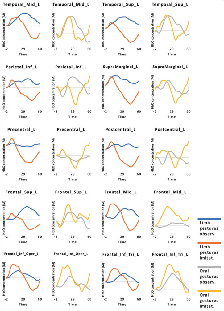
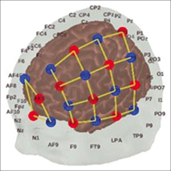
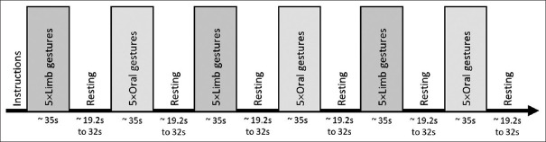
obtained. A continuous‐wave OxyMon fNIRS system (Artinis Medical Systems, Netherlands) with 28 active channels and a 10 Hz sampling rate was used. Measured wavelengths were 762 and 845 nm. Based on previous findings, the middle and superior parts of the temporal lobe, inferior and superior parts of the parietal lobe, and superior, middle, and inferior parts of the frontal lobe were selected as regions of interest.[9‐11] Location of optodes was determined using fNIRS Optodes’ Location Decider[12] and the most similar template was selected [Figure 2]. Raw data were processed using Homer3 in MATLAB 2021a (MathWorks, Natick, MA, USA).[13] After the conversion of light intensity signals to an optical density (OD), a bandpass filter of 0.01–0.1 Hz was applied and targeted principle component analysis was performed.[14] Changes in OD were then converted to concentration changes using modified Beer–Lambert Law.[15] Concentration changes within a period of‐2s before stimulus onset to 60s after stimulus onset (2s for baseline, 35s for five stimuli, and 23s for return to baseline) were averaged to obtain the hemodynamic response functions (HRF) during the task. Next, the HRF from channels within one region of interest (ROI) was averaged. Figures 3 and 4 demonstrate the HRF during the task. Overall, this experiment suggests that fNIRS can be used to study apraxia, especially in elderly patients with neurodegenerative diseases.
功能性近红外光谱在阿尔茨海默病失调症研究中的应用:概念验证实验。
本文章由计算机程序翻译,如有差异,请以英文原文为准。
求助全文
约1分钟内获得全文
求助全文
来源期刊

Journal of Medical Signals & Sensors
ENGINEERING, BIOMEDICAL-
CiteScore
2.30
自引率
0.00%
发文量
53
审稿时长
33 weeks
期刊介绍:
JMSS is an interdisciplinary journal that incorporates all aspects of the biomedical engineering including bioelectrics, bioinformatics, medical physics, health technology assessment, etc. Subject areas covered by the journal include: - Bioelectric: Bioinstruments Biosensors Modeling Biomedical signal processing Medical image analysis and processing Medical imaging devices Control of biological systems Neuromuscular systems Cognitive sciences Telemedicine Robotic Medical ultrasonography Bioelectromagnetics Electrophysiology Cell tracking - Bioinformatics and medical informatics: Analysis of biological data Data mining Stochastic modeling Computational genomics Artificial intelligence & fuzzy Applications Medical softwares Bioalgorithms Electronic health - Biophysics and medical physics: Computed tomography Radiation therapy Laser therapy - Education in biomedical engineering - Health technology assessment - Standard in biomedical engineering.
 求助内容:
求助内容: 应助结果提醒方式:


