{"title":"Preoperative sonographic assessment of a leiomyoma of the round ligament.","authors":"Francesca Arezzo, Gerardo Cazzato, Vera Loizzi, Viviana Cataldo, Michele Mongelli, Claudio Lombardi, Gennaro Cormio","doi":"10.23750/abm.v93iS1.11717","DOIUrl":null,"url":null,"abstract":"We report a case of leiomyoma of the round ligament in a 59-year-old woman, with suspicion of ovarian cancer during gynaecological routine examination. Transvaginal ultrasound showed a heterogeneous solid pelvic mass in the left adnexal area but during the evaluation of its anatomical relation, both ovaries appeared to be regular and the neoformation was separated from the uterus. Surgical and pathological examination revealed well-defined solid mass arising from the left round ligament of the uterus identified as leiomyoma with myxoid degeneration. Leiomyomas of the Round Ligament of the uterus are very rare tumors and they may arise as inguinal, pelvic or vulvar masses miming an inguinal hernia, a lymphadenopathy or a solid adnexal mass. The preoperative sonographic assessment is essential to perform a correct differential diagnosis and provide a right management of the case.","PeriodicalId":93849,"journal":{"name":"Acta bio-medica : Atenei Parmensis","volume":"93 S1","pages":"e2022125"},"PeriodicalIF":0.0000,"publicationDate":"2022-06-07","publicationTypes":"Journal Article","fieldsOfStudy":null,"isOpenAccess":false,"openAccessPdf":"https://ftp.ncbi.nlm.nih.gov/pub/pmc/oa_pdf/b2/16/ACTA-93-125.PMC10510981.pdf","citationCount":"0","resultStr":null,"platform":"Semanticscholar","paperid":null,"PeriodicalName":"Acta bio-medica : Atenei Parmensis","FirstCategoryId":"1085","ListUrlMain":"https://doi.org/10.23750/abm.v93iS1.11717","RegionNum":0,"RegionCategory":null,"ArticlePicture":[],"TitleCN":null,"AbstractTextCN":null,"PMCID":null,"EPubDate":"","PubModel":"","JCR":"","JCRName":"","Score":null,"Total":0}
引用次数: 0
Abstract
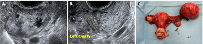
We report a case of leiomyoma of the round ligament in a 59-year-old woman, with suspicion of ovarian cancer during gynaecological routine examination. Transvaginal ultrasound showed a heterogeneous solid pelvic mass in the left adnexal area but during the evaluation of its anatomical relation, both ovaries appeared to be regular and the neoformation was separated from the uterus. Surgical and pathological examination revealed well-defined solid mass arising from the left round ligament of the uterus identified as leiomyoma with myxoid degeneration. Leiomyomas of the Round Ligament of the uterus are very rare tumors and they may arise as inguinal, pelvic or vulvar masses miming an inguinal hernia, a lymphadenopathy or a solid adnexal mass. The preoperative sonographic assessment is essential to perform a correct differential diagnosis and provide a right management of the case.

 求助内容:
求助内容: 应助结果提醒方式:


