Recombinant adenovirus causes prolonged mobilization of macrophages in the anterior chambers of mice.
IF 1.8 3区 医学Q4 BIOCHEMISTRY & MOLECULAR BIOLOGY
Molecular VisionPub Date : 2021-12-28eCollection Date: 2021-01-01
Kacie J Meyer, Danielle Pellack, Adam Hedberg-Buenz, Nicholas Pomernackas, Dana Soukup, Kai Wang, John H Fingert, Michael G Anderson
{"title":"Recombinant adenovirus causes prolonged mobilization of macrophages in the anterior chambers of mice.","authors":"Kacie J Meyer, Danielle Pellack, Adam Hedberg-Buenz, Nicholas Pomernackas, Dana Soukup, Kai Wang, John H Fingert, Michael G Anderson","doi":"","DOIUrl":null,"url":null,"abstract":"<p><strong>Purpose: </strong>Ocular tissues of mice have been studied in many ways using replication-deficient species C type 5 adenovirus (Ad5) as a tool for manipulating gene expression. Whereas refinements to injection protocols and tropism have led to several advances in targeting cells of interest, there remains a relative lack of information concerning how Ad5 may influence other ocular cell types capable of confounding experimental interpretation. Here, a slit lamp is used to thoroughly photodocument the sequelae of intraocular Ad5 injections over time in mice, with attention to potentially confounding indices of inflammation.</p><p><strong>Methods: </strong>A cohort of C57BL/6J mice was randomly split into three groups (Virus, receiving unilateral intracameral injection with 5×10<sup>7</sup> plaque-forming units (pfu) of a cargo-less Ad5 construct; Saline, receiving unilateral balanced salt solution injection; and Naïve, receiving no injections). From this initial experiment, a total of 52 eyes from 26 mice were photodocumented via slit lamp at four time points (baseline and 1, 3, and 10 weeks following initiation of the experiment) by an observer masked to treatments and other parameters of the experimental design. Following the last in vivo exam, tissues were collected. Based on the slit-lamp data, tissues were studied via immunostaining with the macrophage marker F4/80. Subsequently, three iterations of the original experiment were performed with otherwise identical experimental parameters testing the effect of age, intravitreal injection, and A195 buffer, adding slit-lamp photodocumentation of an additional 32 eyes from 16 mice.</p><p><strong>Results: </strong>The masked investigator could use the sequential images from each mouse in the initial experiment to assign each mouse to its correct treatment group with near perfect fidelity. Virus-injected eyes were characterized by corneal damage indicative of intraocular injection and a prolonged mobilization of clump cells on the surface of the iris. Saline-injected eyes had only transient corneal opacities indicative of intraocular injections, and Naïve eyes remained normal. Immunostaining with F4/80 was consistent with ascribing the clump cells visualized via slit-lamp imaging as a type of macrophage. Experimental iterations using Ad5 indicate that all virus-injected eyes had the distinguishing feature of a prolonged presence of clump cells on the surface of the iris regardless of injection site. Mice receiving an intraocular injection of Ad5 at an advanced age displayed a protracted course of corneal cloudiness that prevented detailed visualization of the iris at the last time point.</p><p><strong>Conclusions: </strong>Because the eye is often considered an \"immune privileged site,\" we suspect that several studies have neglected to consider that the presence of Ad5 in the eye might evoke strong reactions from the innate immune system. Ad5 injection caused a sustained mobilization of clump cells-that is, macrophages. This change is likely a consequence of either direct macrophage transduction or a secondary response to cytokines produced locally by other transduced cells. Regardless of how these cells were altered, the important implication is that the adenovirus led to long-lasting changes in the environment of the anterior chamber. Thus, these findings describe a caveat of Ad5-mediated studies involving macrophage mobilization, which we encourage groups to use as a bioassay in their experiments and consider in interpretation of their ongoing experiments using adenoviruses.</p>","PeriodicalId":18866,"journal":{"name":"Molecular Vision","volume":"27 ","pages":"741-756"},"PeriodicalIF":1.8000,"publicationDate":"2021-12-28","publicationTypes":"Journal Article","fieldsOfStudy":null,"isOpenAccess":false,"openAccessPdf":"https://ftp.ncbi.nlm.nih.gov/pub/pmc/oa_pdf/fb/c6/mv-v27-741.PMC8763664.pdf","citationCount":"0","resultStr":null,"platform":"Semanticscholar","paperid":null,"PeriodicalName":"Molecular Vision","FirstCategoryId":"3","ListUrlMain":"","RegionNum":3,"RegionCategory":"医学","ArticlePicture":[],"TitleCN":null,"AbstractTextCN":null,"PMCID":null,"EPubDate":"2021/1/1 0:00:00","PubModel":"eCollection","JCR":"Q4","JCRName":"BIOCHEMISTRY & MOLECULAR BIOLOGY","Score":null,"Total":0}
引用次数: 0
Abstract
Purpose: Ocular tissues of mice have been studied in many ways using replication-deficient species C type 5 adenovirus (Ad5) as a tool for manipulating gene expression. Whereas refinements to injection protocols and tropism have led to several advances in targeting cells of interest, there remains a relative lack of information concerning how Ad5 may influence other ocular cell types capable of confounding experimental interpretation. Here, a slit lamp is used to thoroughly photodocument the sequelae of intraocular Ad5 injections over time in mice, with attention to potentially confounding indices of inflammation.
Methods: A cohort of C57BL/6J mice was randomly split into three groups (Virus, receiving unilateral intracameral injection with 5×10⁷ plaque-forming units (pfu) of a cargo-less Ad5 construct; Saline, receiving unilateral balanced salt solution injection; and Naïve, receiving no injections). From this initial experiment, a total of 52 eyes from 26 mice were photodocumented via slit lamp at four time points (baseline and 1, 3, and 10 weeks following initiation of the experiment) by an observer masked to treatments and other parameters of the experimental design. Following the last in vivo exam, tissues were collected. Based on the slit-lamp data, tissues were studied via immunostaining with the macrophage marker F4/80. Subsequently, three iterations of the original experiment were performed with otherwise identical experimental parameters testing the effect of age, intravitreal injection, and A195 buffer, adding slit-lamp photodocumentation of an additional 32 eyes from 16 mice.
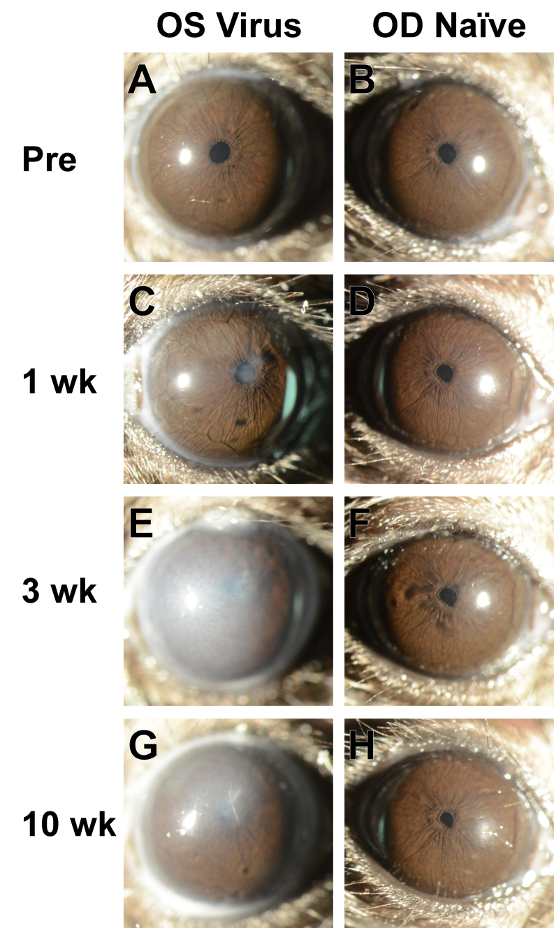
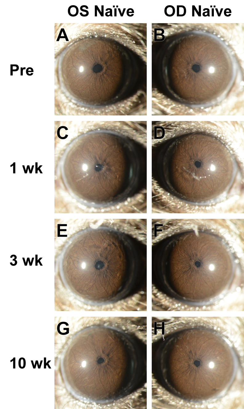
Results: The masked investigator could use the sequential images from each mouse in the initial experiment to assign each mouse to its correct treatment group with near perfect fidelity. Virus-injected eyes were characterized by corneal damage indicative of intraocular injection and a prolonged mobilization of clump cells on the surface of the iris. Saline-injected eyes had only transient corneal opacities indicative of intraocular injections, and Naïve eyes remained normal. Immunostaining with F4/80 was consistent with ascribing the clump cells visualized via slit-lamp imaging as a type of macrophage. Experimental iterations using Ad5 indicate that all virus-injected eyes had the distinguishing feature of a prolonged presence of clump cells on the surface of the iris regardless of injection site. Mice receiving an intraocular injection of Ad5 at an advanced age displayed a protracted course of corneal cloudiness that prevented detailed visualization of the iris at the last time point.
Conclusions: Because the eye is often considered an "immune privileged site," we suspect that several studies have neglected to consider that the presence of Ad5 in the eye might evoke strong reactions from the innate immune system. Ad5 injection caused a sustained mobilization of clump cells-that is, macrophages. This change is likely a consequence of either direct macrophage transduction or a secondary response to cytokines produced locally by other transduced cells. Regardless of how these cells were altered, the important implication is that the adenovirus led to long-lasting changes in the environment of the anterior chamber. Thus, these findings describe a caveat of Ad5-mediated studies involving macrophage mobilization, which we encourage groups to use as a bioassay in their experiments and consider in interpretation of their ongoing experiments using adenoviruses.
期刊介绍:
Molecular Vision is a peer-reviewed journal dedicated to the dissemination of research results in molecular biology, cell biology, and the genetics of the visual system (ocular and cortical).
Molecular Vision publishes articles presenting original research that has not previously been published and comprehensive articles reviewing the current status of a particular field or topic. Submissions to Molecular Vision are subjected to rigorous peer review. Molecular Vision does NOT publish preprints.
For authors, Molecular Vision provides a rapid means of communicating important results. Access to Molecular Vision is free and unrestricted, allowing the widest possible audience for your article. Digital publishing allows you to use color images freely (and without fees). Additionally, you may publish animations, sounds, or other supplementary information that clarifies or supports your article. Each of the authors of an article may also list an electronic mail address (which will be updated upon request) to give interested readers easy access to authors.

 求助内容:
求助内容: 应助结果提醒方式:


