{"title":"Investigation of allosteric coupling in human β2-adrenergic receptor in the presence of intracellular loop 3","authors":"Canan Ozgur, Pemra Doruker, E. Demet Akten","doi":"10.1186/s12900-016-0061-9","DOIUrl":null,"url":null,"abstract":"<p>This study investigates the allosteric coupling that exists between the intra- and extracellular parts of human β<sub>2</sub>-adrenergic receptor (β<sub>2</sub>-AR), in the presence of the intracellular loop 3 (ICL3), which is missing in all crystallographic experiments and most of the simulation studies reported so far. Our recent 1?μs long MD run has revealed a transition to the so-called <i>very inactive</i> state of the receptor, in which ICL3 packed under the G protein’s binding cavity and completely blocked its accessibility to G protein. Simultaneously, an outward tilt of transmembrane helix 5 (TM5) caused an expansion of the extracellular ligand-binding site. In the current study, we performed independent runs with a total duration of 4?μs to further investigate the <i>very inactive</i> state with packed ICL3 and the allosteric coupling event (three unrestrained runs and five runs with bond restraints at the ligand-binding site).</p><p>In all three independent unrestrained runs (each 500?ns long), ICL3 preserved its initially packed/closed conformation within the studied time frame, suggesting an inhibition of the receptor’s activity. Specific bond restraints were later imposed between some key residues at the ligand-binding site, which have been experimentally determined to interact with the ligand. Restraining the binding site region to an open state facilitated ICL3 closure, whereas a relatively constrained/closed binding site hindered ICL3 packing. However, the reverse operation, i.e. opening of the packed ICL3, could not be realized by restraining the binding site region to a closed state. Thus, any attempt failed to free the ICL3 from its locked state due to the presence of persistent hydrogen bonds.</p><p>Overall, our simulations indicated that starting with <i>very inactive</i> states, the receptor stayed almost irreversibly inhibited, which in turn decreased the overall mobility of the receptor. Bond restraints which represented the geometric restrictions caused by ligands of various sizes when bound at the ligand-binding site, induced the expected conformational changes in TM5, TM6 and consequently, ICL3. Still, once ICL3 was packed, the allosteric coupling became ineffective due to strong hydrogen bonds connecting ICL3 to the core of the receptor.</p>","PeriodicalId":51240,"journal":{"name":"BMC Structural Biology","volume":"16 1","pages":""},"PeriodicalIF":0.0000,"publicationDate":"2016-07-02","publicationTypes":"Journal Article","fieldsOfStudy":null,"isOpenAccess":false,"openAccessPdf":"https://sci-hub-pdf.com/10.1186/s12900-016-0061-9","citationCount":"15","resultStr":null,"platform":"Semanticscholar","paperid":null,"PeriodicalName":"BMC Structural Biology","FirstCategoryId":"1085","ListUrlMain":"https://link.springer.com/article/10.1186/s12900-016-0061-9","RegionNum":0,"RegionCategory":null,"ArticlePicture":[],"TitleCN":null,"AbstractTextCN":null,"PMCID":null,"EPubDate":"","PubModel":"","JCR":"Q3","JCRName":"Biochemistry, Genetics and Molecular Biology","Score":null,"Total":0}
引用次数: 15
Abstract
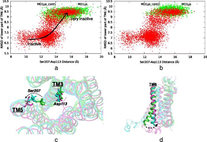
This study investigates the allosteric coupling that exists between the intra- and extracellular parts of human β2-adrenergic receptor (β2-AR), in the presence of the intracellular loop 3 (ICL3), which is missing in all crystallographic experiments and most of the simulation studies reported so far. Our recent 1?μs long MD run has revealed a transition to the so-called very inactive state of the receptor, in which ICL3 packed under the G protein’s binding cavity and completely blocked its accessibility to G protein. Simultaneously, an outward tilt of transmembrane helix 5 (TM5) caused an expansion of the extracellular ligand-binding site. In the current study, we performed independent runs with a total duration of 4?μs to further investigate the very inactive state with packed ICL3 and the allosteric coupling event (three unrestrained runs and five runs with bond restraints at the ligand-binding site).
In all three independent unrestrained runs (each 500?ns long), ICL3 preserved its initially packed/closed conformation within the studied time frame, suggesting an inhibition of the receptor’s activity. Specific bond restraints were later imposed between some key residues at the ligand-binding site, which have been experimentally determined to interact with the ligand. Restraining the binding site region to an open state facilitated ICL3 closure, whereas a relatively constrained/closed binding site hindered ICL3 packing. However, the reverse operation, i.e. opening of the packed ICL3, could not be realized by restraining the binding site region to a closed state. Thus, any attempt failed to free the ICL3 from its locked state due to the presence of persistent hydrogen bonds.
Overall, our simulations indicated that starting with very inactive states, the receptor stayed almost irreversibly inhibited, which in turn decreased the overall mobility of the receptor. Bond restraints which represented the geometric restrictions caused by ligands of various sizes when bound at the ligand-binding site, induced the expected conformational changes in TM5, TM6 and consequently, ICL3. Still, once ICL3 was packed, the allosteric coupling became ineffective due to strong hydrogen bonds connecting ICL3 to the core of the receptor.
期刊介绍:
BMC Structural Biology is an open access, peer-reviewed journal that considers articles on investigations into the structure of biological macromolecules, including solving structures, structural and functional analyses, and computational modeling.


 求助内容:
 应助结果提醒方式:


 京公网安备 11010802042870号