Ching-Yee Oliver Wong, James Gannon, Jeffrey Bong, Christiana O Wong, Gopal B Saha
{"title":"Computer-assisted lateralization of unilateral temporal lobe epilepsy using Z-score parametric F-18 FDG PET images.","authors":"Ching-Yee Oliver Wong, James Gannon, Jeffrey Bong, Christiana O Wong, Gopal B Saha","doi":"10.1186/1471-2385-7-5","DOIUrl":null,"url":null,"abstract":"<p><strong>Background: </strong>To evaluate the use of unbiased computer-assisted lateralization of temporal lobe epilepsy (TLE) by z-score parametric PET imaging (ZPET).</p><p><strong>Methods: </strong>38 patients with histologically proven unilateral TLE due to pure hippocampal sclerosis, referred for pre-surgical PET evaluation of intractable seizure over a 5-year period, were included. The F-18 FDG images were oriented along temporal long axis and then transformed into ZPET images on a voxel by voxel basis. Multiple regions of interests (21 in total) were placed on cortical, subcortical and cerebellar structures on twenty-eight out of 38 patients with totally seizure-free (class I) outcome. Paired t-tests with Bonferroni correction were used to determine the location of the most asymmetric regions as variables for subsequent discriminant analysis of the entire group of the patients.</p><p><strong>Results: </strong>The computer program identified the anterior half of the temporal lobe (p < 0.0005) and thalami (p = 0.021) as the most asymmetric regions in TLE patients with Class I outcome. Discriminant analysis using z-scores from a total of 8 ROIs (in 4 pairs) on these structures correctly lateralized thirty-seven out of 38 (97%) patients (sensitivity = 94%; specificity = 100%). The only false localization came from a patient with equivocal z-scores on the temporal lobes and this patient turned out to have poor outcome.</p><p><strong>Conclusion: </strong>The computer-assisted lateralization of TLE using ZPET provides an accurate, fast and objective way of seizure evaluation.</p>","PeriodicalId":80684,"journal":{"name":"BMC nuclear medicine","volume":" ","pages":"5"},"PeriodicalIF":0.0000,"publicationDate":"2007-11-02","publicationTypes":"Journal Article","fieldsOfStudy":null,"isOpenAccess":false,"openAccessPdf":"https://sci-hub-pdf.com/10.1186/1471-2385-7-5","citationCount":"1","resultStr":null,"platform":"Semanticscholar","paperid":null,"PeriodicalName":"BMC nuclear medicine","FirstCategoryId":"1085","ListUrlMain":"https://doi.org/10.1186/1471-2385-7-5","RegionNum":0,"RegionCategory":null,"ArticlePicture":[],"TitleCN":null,"AbstractTextCN":null,"PMCID":null,"EPubDate":"","PubModel":"","JCR":"","JCRName":"","Score":null,"Total":0}
引用次数: 1
Abstract
Background: To evaluate the use of unbiased computer-assisted lateralization of temporal lobe epilepsy (TLE) by z-score parametric PET imaging (ZPET).
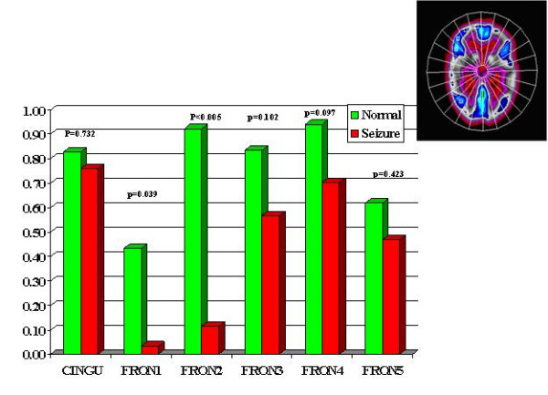
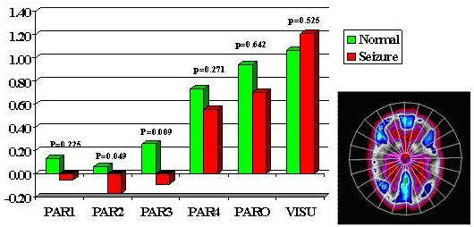
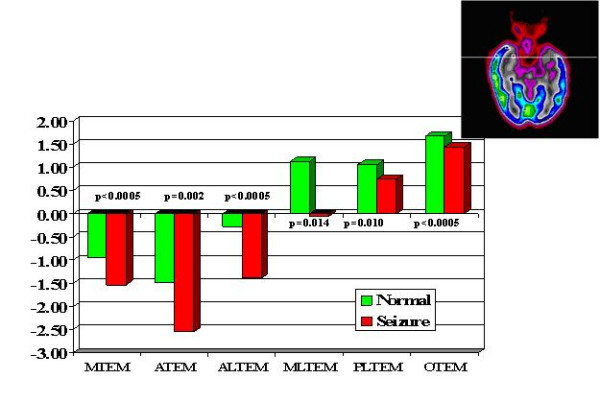
Methods: 38 patients with histologically proven unilateral TLE due to pure hippocampal sclerosis, referred for pre-surgical PET evaluation of intractable seizure over a 5-year period, were included. The F-18 FDG images were oriented along temporal long axis and then transformed into ZPET images on a voxel by voxel basis. Multiple regions of interests (21 in total) were placed on cortical, subcortical and cerebellar structures on twenty-eight out of 38 patients with totally seizure-free (class I) outcome. Paired t-tests with Bonferroni correction were used to determine the location of the most asymmetric regions as variables for subsequent discriminant analysis of the entire group of the patients.
Results: The computer program identified the anterior half of the temporal lobe (p < 0.0005) and thalami (p = 0.021) as the most asymmetric regions in TLE patients with Class I outcome. Discriminant analysis using z-scores from a total of 8 ROIs (in 4 pairs) on these structures correctly lateralized thirty-seven out of 38 (97%) patients (sensitivity = 94%; specificity = 100%). The only false localization came from a patient with equivocal z-scores on the temporal lobes and this patient turned out to have poor outcome.
Conclusion: The computer-assisted lateralization of TLE using ZPET provides an accurate, fast and objective way of seizure evaluation.

 求助内容:
求助内容: 应助结果提醒方式:


