Jennifer Sartor-Pfeiffer, Mirjam Lingel, Maria-Ioanna Stefanou, Tobias Lindig, Benjamin Bender, Sven Poli, Ulf Ziemann, Andreas Fritsche, Katharina Feil, Annerose Mengel
{"title":"Regional computed tomography perfusion deficits in patients with hypoglycemia: two case reports.","authors":"Jennifer Sartor-Pfeiffer, Mirjam Lingel, Maria-Ioanna Stefanou, Tobias Lindig, Benjamin Bender, Sven Poli, Ulf Ziemann, Andreas Fritsche, Katharina Feil, Annerose Mengel","doi":"10.1186/s42466-022-00201-z","DOIUrl":null,"url":null,"abstract":"<p><strong>Background: </strong>Hypoglycemia in patients with diabetes mellitus, particularly type 1 can mimic acute ischemic stroke by causing focal neurological deficits. In acute ischemic stroke, the interpretation of emergency imaging including computed tomography with angiography and perfusion is crucial to guide revascularizing therapy including intravenous thrombolysis. However, different metabolic abnormalities and stroke mimics can cause focal hypoperfusion.</p><p><strong>Methods: </strong>We describe two type 1 diabetes patients presenting with acute focal neurological deficits and hypoglycemia, who underwent multimodal computed tomography and follow-up imaging.</p><p><strong>Case presentation: </strong>Patient 1, a 20-year-old man presented with aphasia and interstitial glucose level of 54 mg/dl. Patient 2, a 77-year-old man presented with aphasia, mild right-sided brachiofacial paresis and interstitial glucose level of 83 mg/dl. On brain imaging, no acute infarct signs were noted. Yet, both had focal left hemispheric cerebral hypoperfusion without large-vessel occlusion or stenosis. Due to persistent symptoms after normalization of blood glucose and despite a perfusion imaging pattern that was interpretated as non-typical for ischemia, both patients underwent thrombolysis without any complications.</p><p><strong>Conclusion: </strong>Computed tomography perfusion might help to discriminate hypoglycemia with focal neurological signs from acute stroke, but further evidence is needed.</p>","PeriodicalId":19169,"journal":{"name":"Neurological Research and Practice","volume":" ","pages":"36"},"PeriodicalIF":0.0000,"publicationDate":"2022-08-22","publicationTypes":"Journal Article","fieldsOfStudy":null,"isOpenAccess":false,"openAccessPdf":"https://www.ncbi.nlm.nih.gov/pmc/articles/PMC9394021/pdf/","citationCount":"1","resultStr":null,"platform":"Semanticscholar","paperid":null,"PeriodicalName":"Neurological Research and Practice","FirstCategoryId":"1085","ListUrlMain":"https://doi.org/10.1186/s42466-022-00201-z","RegionNum":0,"RegionCategory":null,"ArticlePicture":[],"TitleCN":null,"AbstractTextCN":null,"PMCID":null,"EPubDate":"","PubModel":"","JCR":"","JCRName":"","Score":null,"Total":0}
引用次数: 1
Abstract
Background: Hypoglycemia in patients with diabetes mellitus, particularly type 1 can mimic acute ischemic stroke by causing focal neurological deficits. In acute ischemic stroke, the interpretation of emergency imaging including computed tomography with angiography and perfusion is crucial to guide revascularizing therapy including intravenous thrombolysis. However, different metabolic abnormalities and stroke mimics can cause focal hypoperfusion.
Methods: We describe two type 1 diabetes patients presenting with acute focal neurological deficits and hypoglycemia, who underwent multimodal computed tomography and follow-up imaging.
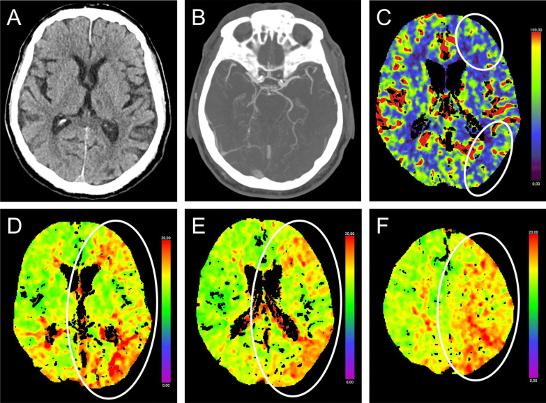
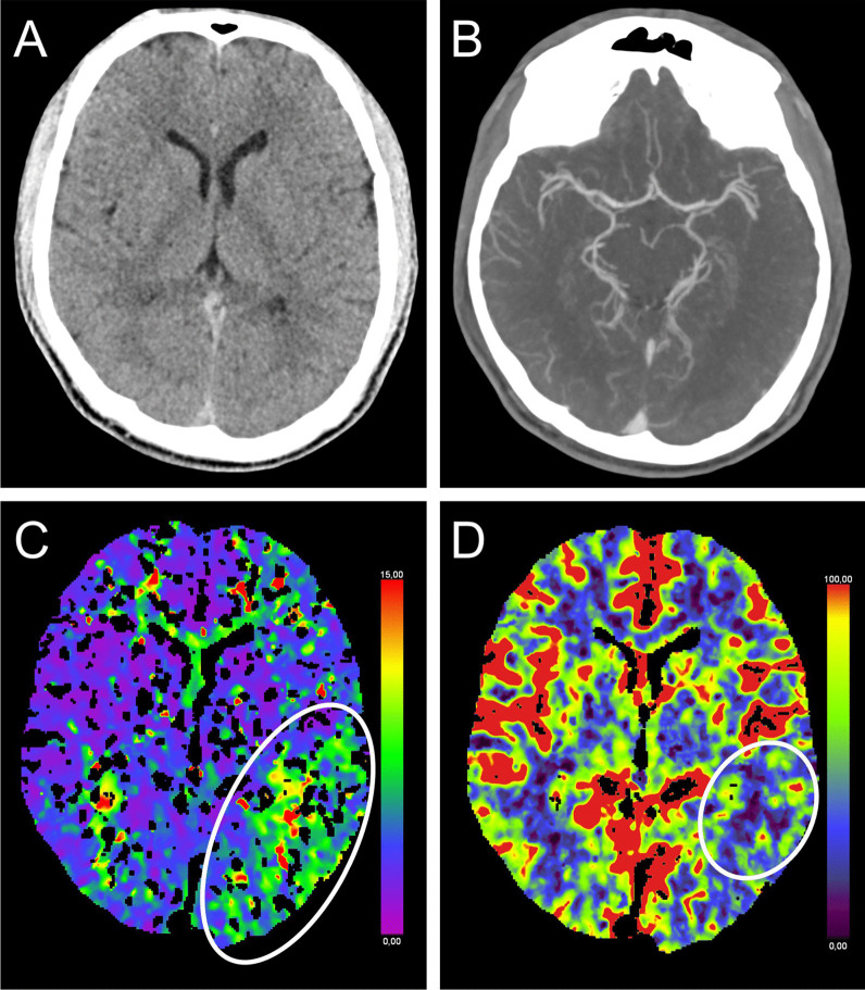
Case presentation: Patient 1, a 20-year-old man presented with aphasia and interstitial glucose level of 54 mg/dl. Patient 2, a 77-year-old man presented with aphasia, mild right-sided brachiofacial paresis and interstitial glucose level of 83 mg/dl. On brain imaging, no acute infarct signs were noted. Yet, both had focal left hemispheric cerebral hypoperfusion without large-vessel occlusion or stenosis. Due to persistent symptoms after normalization of blood glucose and despite a perfusion imaging pattern that was interpretated as non-typical for ischemia, both patients underwent thrombolysis without any complications.
Conclusion: Computed tomography perfusion might help to discriminate hypoglycemia with focal neurological signs from acute stroke, but further evidence is needed.

 求助内容:
求助内容: 应助结果提醒方式:


