Sarah A. Pfaff, Xuan Wang, Edward R. Wagner, Liza A. Wilson, Sarah N. Kiemle, Daniel J. Cosgrove
{"title":"Detecting the orientation of newly-deposited crystalline cellulose with fluorescent CBM3","authors":"Sarah A. Pfaff, Xuan Wang, Edward R. Wagner, Liza A. Wilson, Sarah N. Kiemle, Daniel J. Cosgrove","doi":"10.1016/j.tcsw.2022.100089","DOIUrl":null,"url":null,"abstract":"<div><p>Cellulose microfibril patterning influences many of the mechanical attributes of plant cell walls. We developed a simple, fluorescence microscopy-based method to detect the orientation of newly-synthesized cellulose microfibrils in epidermal peels of onion and Arabidopsis. It is based on Alexa Fluor 488-tagged carbohydrate binding module 3a (CBM3a) from <em>Clostridium thermocellum</em> which displayed a nearly 4-fold greater binding to cell walls at pH 5.5 compared with pH 8. Binding to isolated cellulose did not display this pH dependence. At pH 7.5 fibrillar patterns at the surface of the epidermal peels were visible, corresponding to the directionality of surface cellulose microfibrils, as verified by atomic force microscopy. The fibrillar pattern was not visible as the labeling intensity increased at lower pH. The pH of greatest cell wall labeling corresponds to the isoelectric point of CBM3a, suggesting that electrostatic forces limit CBM3a penetration into the wall. Consistent with this, digestion of the wall with pectate lyase to remove homogalacturonan increased labeling intensity. We conclude that electrostatic interactions strongly influence labeling of cell walls with CBM3 and potentially other proteins, holding implications for any work that relies on penetration of protein probes such as CBMs, antibodies, or enzymes into charged polymeric substrates.</p></div>","PeriodicalId":36539,"journal":{"name":"Cell Surface","volume":"8 ","pages":"Article 100089"},"PeriodicalIF":6.2000,"publicationDate":"2022-12-01","publicationTypes":"Journal Article","fieldsOfStudy":null,"isOpenAccess":false,"openAccessPdf":"https://ftp.ncbi.nlm.nih.gov/pub/pmc/oa_pdf/5f/b7/main.PMC9678952.pdf","citationCount":"0","resultStr":null,"platform":"Semanticscholar","paperid":null,"PeriodicalName":"Cell Surface","FirstCategoryId":"1085","ListUrlMain":"https://www.sciencedirect.com/science/article/pii/S2468233022000184","RegionNum":0,"RegionCategory":null,"ArticlePicture":[],"TitleCN":null,"AbstractTextCN":null,"PMCID":null,"EPubDate":"","PubModel":"","JCR":"Q1","JCRName":"Immunology and Microbiology","Score":null,"Total":0}
引用次数: 0
Abstract
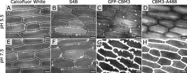
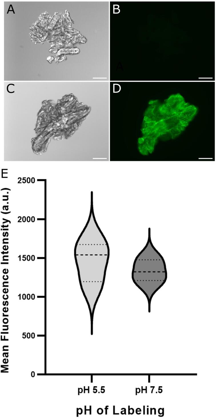
Cellulose microfibril patterning influences many of the mechanical attributes of plant cell walls. We developed a simple, fluorescence microscopy-based method to detect the orientation of newly-synthesized cellulose microfibrils in epidermal peels of onion and Arabidopsis. It is based on Alexa Fluor 488-tagged carbohydrate binding module 3a (CBM3a) from Clostridium thermocellum which displayed a nearly 4-fold greater binding to cell walls at pH 5.5 compared with pH 8. Binding to isolated cellulose did not display this pH dependence. At pH 7.5 fibrillar patterns at the surface of the epidermal peels were visible, corresponding to the directionality of surface cellulose microfibrils, as verified by atomic force microscopy. The fibrillar pattern was not visible as the labeling intensity increased at lower pH. The pH of greatest cell wall labeling corresponds to the isoelectric point of CBM3a, suggesting that electrostatic forces limit CBM3a penetration into the wall. Consistent with this, digestion of the wall with pectate lyase to remove homogalacturonan increased labeling intensity. We conclude that electrostatic interactions strongly influence labeling of cell walls with CBM3 and potentially other proteins, holding implications for any work that relies on penetration of protein probes such as CBMs, antibodies, or enzymes into charged polymeric substrates.

 求助内容:
求助内容: 应助结果提醒方式:


