Albert Stezin, Venkateswara Reddy Reddam, Shantala Hegde, Ravi Yadav, Jitender Saini, Pramod Kumar Pal
{"title":"Morphometric mapping of the macrostructural abnormalities of midsagittal corpus callosum in Wilson's disease.","authors":"Albert Stezin, Venkateswara Reddy Reddam, Shantala Hegde, Ravi Yadav, Jitender Saini, Pramod Kumar Pal","doi":"10.4103/AOMD.AOMD_41_20","DOIUrl":null,"url":null,"abstract":"<p><strong>Background and purpose: </strong>The corpus callosum (CC) consists of topographically arranged white matter (WM) fibers. Previous studies have indicated the CC to be discretely involved in WD. In this study, we strived to characterize the macrostructural properties of the CC using midsagittal cross-sectional area and thickness profile measurements.</p><p><strong>Materials and methods: </strong>This study was performed using archived magnetic resonance imaging (MRI) scans of 14 patients with WD and 14 age- and gender-matched healthy controls. Using an automated software pipeline for morphometric profiling, the midsagittal CC was segmented into five sub-regions (CC<sub>1-5</sub>) according to the Hofer-Frahm scheme. The mean thickness and area of different CC segments and their clinical and cognitive correlates were identified.</p><p><strong>Results: </strong>The mean area was significantly different only in CC<sub>2</sub> segment (94.2 ± 25.5 vs. 118.6 ± 19.7 mm<sup>2</sup>, corrected <i>P</i> < 0.05). The mean thickness was significantly different in CC<sub>1</sub> (5.06 ± 1.15 vs. 6.93 ± 0.89 mm, corrected <i>P</i> < 0.05), CC<sub>2</sub> (3.73 ± 0.96 vs. 4.87 ± 1.01 mm, corrected <i>P</i> < 0.05), and CC<sub>3</sub> segments (3.42 ± 0.84 vs. 3.94 ± 0.72 mm, corrected <i>P</i> < 0.05). The age at onset of neurological symptoms and MMSE score was significantly correlated with the morphometric changes of CC<sub>1</sub> and CC<sub>2</sub> segments.</p><p><strong>Conclusion: </strong>Morphological changes of the CC are discrete in WD. Morphometric loss of CC was associated with an earlier onset of neurological symptoms and cognitive dysfunction in WD.</p>","PeriodicalId":7973,"journal":{"name":"Annals of Movement Disorders","volume":"4 2","pages":"60-65"},"PeriodicalIF":0.0000,"publicationDate":"2021-05-31","publicationTypes":"Journal Article","fieldsOfStudy":null,"isOpenAccess":false,"openAccessPdf":"https://ftp.ncbi.nlm.nih.gov/pub/pmc/oa_pdf/74/5e/EMS150885.PMC7613241.pdf","citationCount":"0","resultStr":null,"platform":"Semanticscholar","paperid":null,"PeriodicalName":"Annals of Movement Disorders","FirstCategoryId":"1085","ListUrlMain":"https://doi.org/10.4103/AOMD.AOMD_41_20","RegionNum":0,"RegionCategory":null,"ArticlePicture":[],"TitleCN":null,"AbstractTextCN":null,"PMCID":null,"EPubDate":"","PubModel":"","JCR":"Q3","JCRName":"Medicine","Score":null,"Total":0}
引用次数: 0
Abstract
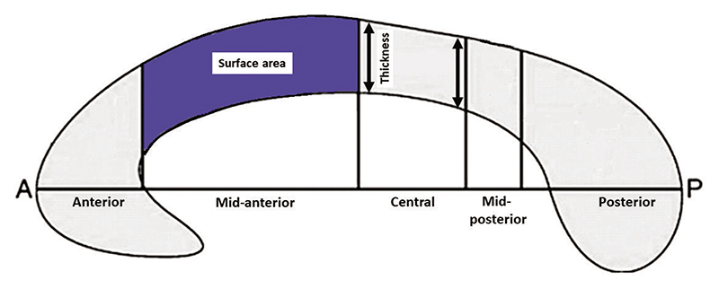
Background and purpose: The corpus callosum (CC) consists of topographically arranged white matter (WM) fibers. Previous studies have indicated the CC to be discretely involved in WD. In this study, we strived to characterize the macrostructural properties of the CC using midsagittal cross-sectional area and thickness profile measurements.
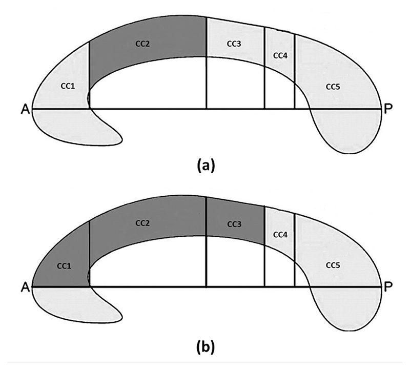
Materials and methods: This study was performed using archived magnetic resonance imaging (MRI) scans of 14 patients with WD and 14 age- and gender-matched healthy controls. Using an automated software pipeline for morphometric profiling, the midsagittal CC was segmented into five sub-regions (CC1-5) according to the Hofer-Frahm scheme. The mean thickness and area of different CC segments and their clinical and cognitive correlates were identified.
Results: The mean area was significantly different only in CC2 segment (94.2 ± 25.5 vs. 118.6 ± 19.7 mm2, corrected P < 0.05). The mean thickness was significantly different in CC1 (5.06 ± 1.15 vs. 6.93 ± 0.89 mm, corrected P < 0.05), CC2 (3.73 ± 0.96 vs. 4.87 ± 1.01 mm, corrected P < 0.05), and CC3 segments (3.42 ± 0.84 vs. 3.94 ± 0.72 mm, corrected P < 0.05). The age at onset of neurological symptoms and MMSE score was significantly correlated with the morphometric changes of CC1 and CC2 segments.
Conclusion: Morphological changes of the CC are discrete in WD. Morphometric loss of CC was associated with an earlier onset of neurological symptoms and cognitive dysfunction in WD.

 求助内容:
求助内容: 应助结果提醒方式:


