Rozemarijn Vliegenthart, Andreas Fouras, Colin Jacobs, Nickolas Papanikolaou
{"title":"Innovations in thoracic imaging: CT, radiomics, AI and x-ray velocimetry.","authors":"Rozemarijn Vliegenthart, Andreas Fouras, Colin Jacobs, Nickolas Papanikolaou","doi":"10.1111/resp.14344","DOIUrl":null,"url":null,"abstract":"<p><p>In recent years, pulmonary imaging has seen enormous progress, with the introduction, validation and implementation of new hardware and software. There is a general trend from mere visual evaluation of radiological images to quantification of abnormalities and biomarkers, and assessment of 'non visual' markers that contribute to establishing diagnosis or prognosis. Important catalysts to these developments in thoracic imaging include new indications (like computed tomography [CT] lung cancer screening) and the COVID-19 pandemic. This review focuses on developments in CT, radiomics, artificial intelligence (AI) and x-ray velocimetry for imaging of the lungs. Recent developments in CT include the potential for ultra-low-dose CT imaging for lung nodules, and the advent of a new generation of CT systems based on photon-counting detector technology. Radiomics has demonstrated potential towards predictive and prognostic tasks particularly in lung cancer, previously not achievable by visual inspection by radiologists, exploiting high dimensional patterns (mostly texture related) on medical imaging data. Deep learning technology has revolutionized the field of AI and as a result, performance of AI algorithms is approaching human performance for an increasing number of specific tasks. X-ray velocimetry integrates x-ray (fluoroscopic) imaging with unique image processing to produce quantitative four dimensional measurement of lung tissue motion, and accurate calculations of lung ventilation.</p>","PeriodicalId":162871,"journal":{"name":"Respirology (Carlton, Vic.)","volume":" ","pages":"818-833"},"PeriodicalIF":0.0000,"publicationDate":"2022-10-01","publicationTypes":"Journal Article","fieldsOfStudy":null,"isOpenAccess":false,"openAccessPdf":"https://www.ncbi.nlm.nih.gov/pmc/articles/PMC9546393/pdf/","citationCount":"14","resultStr":null,"platform":"Semanticscholar","paperid":null,"PeriodicalName":"Respirology (Carlton, Vic.)","FirstCategoryId":"3","ListUrlMain":"https://doi.org/10.1111/resp.14344","RegionNum":0,"RegionCategory":null,"ArticlePicture":[],"TitleCN":null,"AbstractTextCN":null,"PMCID":null,"EPubDate":"2022/8/14 0:00:00","PubModel":"Epub","JCR":"","JCRName":"","Score":null,"Total":0}
引用次数: 14
Abstract
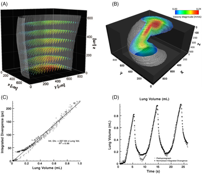
In recent years, pulmonary imaging has seen enormous progress, with the introduction, validation and implementation of new hardware and software. There is a general trend from mere visual evaluation of radiological images to quantification of abnormalities and biomarkers, and assessment of 'non visual' markers that contribute to establishing diagnosis or prognosis. Important catalysts to these developments in thoracic imaging include new indications (like computed tomography [CT] lung cancer screening) and the COVID-19 pandemic. This review focuses on developments in CT, radiomics, artificial intelligence (AI) and x-ray velocimetry for imaging of the lungs. Recent developments in CT include the potential for ultra-low-dose CT imaging for lung nodules, and the advent of a new generation of CT systems based on photon-counting detector technology. Radiomics has demonstrated potential towards predictive and prognostic tasks particularly in lung cancer, previously not achievable by visual inspection by radiologists, exploiting high dimensional patterns (mostly texture related) on medical imaging data. Deep learning technology has revolutionized the field of AI and as a result, performance of AI algorithms is approaching human performance for an increasing number of specific tasks. X-ray velocimetry integrates x-ray (fluoroscopic) imaging with unique image processing to produce quantitative four dimensional measurement of lung tissue motion, and accurate calculations of lung ventilation.

 求助内容:
求助内容: 应助结果提醒方式:


