{"title":"Iatrogenic distal femur fracture following medial femoral supracondylar bone graft harvest: a case report and finite element analysis.","authors":"Sotetsu Sakamoto, Yasunori Hattori, Kazuteru Doi, Hiroki Yamagata, Norihiro Nishida, Takashi Sakai","doi":"10.2185/jrm.2022-032","DOIUrl":null,"url":null,"abstract":"<p><p><b>Objective:</b> This report presents a case of supracondylar femur fracture with finite element analysis and discusses its causes and prevention. <b>Patient and Methods:</b> A 53-year-old man presented with right talar osteonecrosis after osteosynthesis for a talus fracture. A medial femoral condyle-free vascularized bone graft (size, 20 × 12 × 17 mm) from the contralateral femur was performed, including the posteromedial cortical corner. The patient suffered a donor-site supracondylar femoral fracture while standing up from a cross-legged sitting position on the bed on postoperative day 6. The fracture was treated with intramedullary nailing. We analyzed the effects of the location of the bone graft harvest in an intact model using the three-dimensional finite element method (FEM). <b>Results:</b> The talar necrosis and the femur fracture healed. The FEM result revealed that the longitudinal axial pressure had minimal effect on the femur; however, the stress around the bone defect increased with rotation, especially in the posteromedial bone defect model. <b>Conclusion:</b> Harvesting the bone graft should not include the posteromedial corner of the supracondylar femur. The patient should strictly limit the motion of torsional stress, such as standing from a cross-legged sitting position or pivoting turn.</p>","PeriodicalId":73939,"journal":{"name":"Journal of rural medicine : JRM","volume":"17 4","pages":"270-275"},"PeriodicalIF":0.0000,"publicationDate":"2022-10-01","publicationTypes":"Journal Article","fieldsOfStudy":null,"isOpenAccess":false,"openAccessPdf":"https://ftp.ncbi.nlm.nih.gov/pub/pmc/oa_pdf/b7/67/jrm-17-270.PMC9613362.pdf","citationCount":"0","resultStr":null,"platform":"Semanticscholar","paperid":null,"PeriodicalName":"Journal of rural medicine : JRM","FirstCategoryId":"1085","ListUrlMain":"https://doi.org/10.2185/jrm.2022-032","RegionNum":0,"RegionCategory":null,"ArticlePicture":[],"TitleCN":null,"AbstractTextCN":null,"PMCID":null,"EPubDate":"2022/10/22 0:00:00","PubModel":"Epub","JCR":"","JCRName":"","Score":null,"Total":0}
引用次数: 0
Abstract
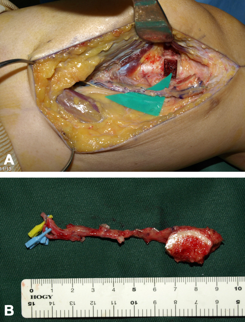
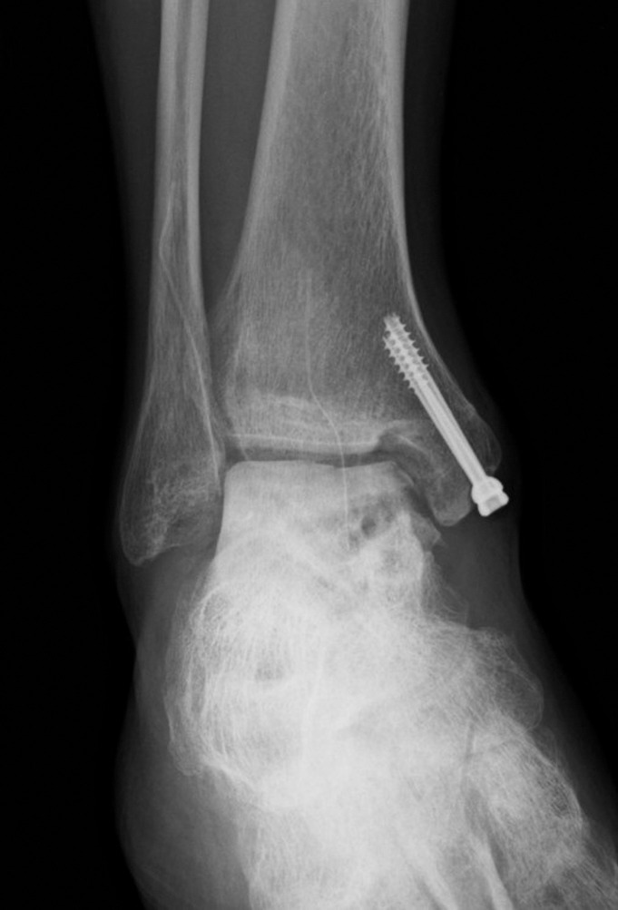
Objective: This report presents a case of supracondylar femur fracture with finite element analysis and discusses its causes and prevention. Patient and Methods: A 53-year-old man presented with right talar osteonecrosis after osteosynthesis for a talus fracture. A medial femoral condyle-free vascularized bone graft (size, 20 × 12 × 17 mm) from the contralateral femur was performed, including the posteromedial cortical corner. The patient suffered a donor-site supracondylar femoral fracture while standing up from a cross-legged sitting position on the bed on postoperative day 6. The fracture was treated with intramedullary nailing. We analyzed the effects of the location of the bone graft harvest in an intact model using the three-dimensional finite element method (FEM). Results: The talar necrosis and the femur fracture healed. The FEM result revealed that the longitudinal axial pressure had minimal effect on the femur; however, the stress around the bone defect increased with rotation, especially in the posteromedial bone defect model. Conclusion: Harvesting the bone graft should not include the posteromedial corner of the supracondylar femur. The patient should strictly limit the motion of torsional stress, such as standing from a cross-legged sitting position or pivoting turn.


 求助内容:
求助内容: 应助结果提醒方式:


