Min Ju Kim, Wonju Hong, Tae Jung Kim, Joungho Han, Yoon-La Choi, Joon Young Choi, Sang Min Lee, Sung Ho Hwang
{"title":"Solitary Pulmonary Capillary Hemangioma: CT and PET-CT Features with Clinicopathologic Correlation.","authors":"Min Ju Kim, Wonju Hong, Tae Jung Kim, Joungho Han, Yoon-La Choi, Joon Young Choi, Sang Min Lee, Sung Ho Hwang","doi":"10.3390/diagnostics12112618","DOIUrl":null,"url":null,"abstract":"<p><p>The aim of this study was to evaluate the CT and PET-CT features of solitary pulmonary capillary hemangioma (SPCH) with clinicopathologic correlations. This retrospective study included 17 patients with histologically proven SPCH from four tertiary institutions. The clinical, pathological and imaging findings of SPCH were reviewed. The CT features assessed included lesion location, size, density, contour, margin, enhancement, presence of air bronchogram, perivascular lucency and pleural retraction, and <sup>18</sup>F-fluorodeoxyglucose uptake on PET-CT. Changes in the size during the follow-up period were also evaluated. Imaging features were correlated with the clinicopathologic findings. The mean age of the patients was 47 years (range 30-60 years). All SPCHs were incidentally detected during screening CT examinations (<i>n</i> = 13, 76%) or during cancer work-up (<i>n</i> = 4, 24%). Most SPCHs appeared as part-solid nodules (<i>n</i> = 15, 88%), the remaining appeared as a pure ground-glass nodule or a pure solid nodule, respectively. Most had smooth contours (<i>n</i> = 16, 94%), while one had a lobulated contour. Nine SPCHs (53%) showed ill-defined margins. Air bronchogram was present in ten (59%) SPCHs, and perivascular lucency in two (12%). All SPCHs exhibited hypoattenuation on contrast-enhanced CT and hypometabolism on PET-CT. During the follow-up period (mean 14.8 ± 17.7 months), the lesions showed no change in size or density in ten SPCHs (59%), decreased or fluctuation in size and density in three (18%). SPCH is often incidentally detected in young and middle-aged adults, commonly as an ill-defined part-solid nodule that may accompany air bronchogram, perivascular lucency, and fluctuation in size or density on CT and hypometabolism on PET-CT.</p>","PeriodicalId":520604,"journal":{"name":"Diagnostics (Basel, Switzerland)","volume":" ","pages":""},"PeriodicalIF":3.3000,"publicationDate":"2022-10-28","publicationTypes":"Journal Article","fieldsOfStudy":null,"isOpenAccess":false,"openAccessPdf":"https://www.ncbi.nlm.nih.gov/pmc/articles/PMC9689303/pdf/","citationCount":"1","resultStr":null,"platform":"Semanticscholar","paperid":null,"PeriodicalName":"Diagnostics (Basel, Switzerland)","FirstCategoryId":"3","ListUrlMain":"https://doi.org/10.3390/diagnostics12112618","RegionNum":0,"RegionCategory":null,"ArticlePicture":[],"TitleCN":null,"AbstractTextCN":null,"PMCID":null,"EPubDate":"","PubModel":"","JCR":"","JCRName":"","Score":null,"Total":0}
引用次数: 1
Abstract
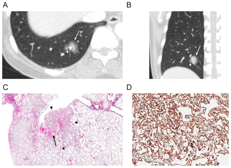
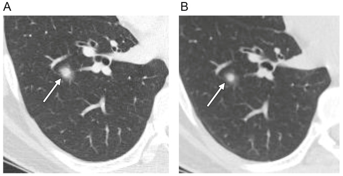
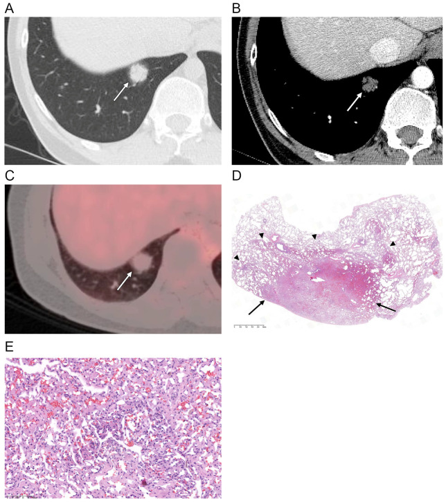
The aim of this study was to evaluate the CT and PET-CT features of solitary pulmonary capillary hemangioma (SPCH) with clinicopathologic correlations. This retrospective study included 17 patients with histologically proven SPCH from four tertiary institutions. The clinical, pathological and imaging findings of SPCH were reviewed. The CT features assessed included lesion location, size, density, contour, margin, enhancement, presence of air bronchogram, perivascular lucency and pleural retraction, and 18F-fluorodeoxyglucose uptake on PET-CT. Changes in the size during the follow-up period were also evaluated. Imaging features were correlated with the clinicopathologic findings. The mean age of the patients was 47 years (range 30-60 years). All SPCHs were incidentally detected during screening CT examinations (n = 13, 76%) or during cancer work-up (n = 4, 24%). Most SPCHs appeared as part-solid nodules (n = 15, 88%), the remaining appeared as a pure ground-glass nodule or a pure solid nodule, respectively. Most had smooth contours (n = 16, 94%), while one had a lobulated contour. Nine SPCHs (53%) showed ill-defined margins. Air bronchogram was present in ten (59%) SPCHs, and perivascular lucency in two (12%). All SPCHs exhibited hypoattenuation on contrast-enhanced CT and hypometabolism on PET-CT. During the follow-up period (mean 14.8 ± 17.7 months), the lesions showed no change in size or density in ten SPCHs (59%), decreased or fluctuation in size and density in three (18%). SPCH is often incidentally detected in young and middle-aged adults, commonly as an ill-defined part-solid nodule that may accompany air bronchogram, perivascular lucency, and fluctuation in size or density on CT and hypometabolism on PET-CT.

 求助内容:
求助内容: 应助结果提醒方式:


