{"title":"A case report of a patient with severe thyroid eye disease.","authors":"Mioara-Laura Macovei, Űnal Azis","doi":"10.22336/rjo.2022.30","DOIUrl":null,"url":null,"abstract":"<p><p><b>Objective:</b> Our aim was to present a rare case of a middle-aged male patient, diagnosed with Graves' orbitopathy, which had an atypical rapid unilateral onset. Initially, the left eye presented exophthalmos, eyelid retraction, corneal ulceration, and pannus formation with an important vascular component due to corneal exposure. The same symptoms developed in the right eye within a short period of time. <b>Methods:</b> A 52-year-old man presented in our department with bilateral proptosis, decrease in visual acuity, and orbital pain, which developed initially in the left eye seven months before the right eye. Slit lamp examination revealed conjunctival hyperemia, purulent discharge, chemosis and inflammation of the caruncle in both eyes. The fluorescein eye stain test was positive due to corneal ulceration with the presence of cells and flare in anterior chamber in the RE (right eye). The LE (left eye) presented a corneal pannus. We documented the changes using a slit lamp biomicroscope, a fundus camera, orbital ultrasonography, and contrast CT (computer tomography) scans. <b>Discussions:</b> The severe Graves' ophthalmopathy represents a challenge both in active or inactive phase. Medical and surgical therapies should be taken into consideration in order to prevent the complications following corneal perforation or optic neuropathy. Also, ophthalmic, and systemic adverse reactions of systemic steroids used in the treatment of Graves' disease are important in the prognosis of the visual outcome. <b>Conclusions:</b> The management of Graves' ophthalmopathy is multidisciplinary and needs a very good therapy adherence in order to achieve a satisfactory prognosis and quality of life.</p>","PeriodicalId":21385,"journal":{"name":"Romanian journal of ophthalmology","volume":"66 2","pages":"153-157"},"PeriodicalIF":0.0000,"publicationDate":"2022-04-01","publicationTypes":"Journal Article","fieldsOfStudy":null,"isOpenAccess":false,"openAccessPdf":"https://www.ncbi.nlm.nih.gov/pmc/articles/PMC9289779/pdf/","citationCount":"3","resultStr":null,"platform":"Semanticscholar","paperid":null,"PeriodicalName":"Romanian journal of ophthalmology","FirstCategoryId":"1085","ListUrlMain":"https://doi.org/10.22336/rjo.2022.30","RegionNum":0,"RegionCategory":null,"ArticlePicture":[],"TitleCN":null,"AbstractTextCN":null,"PMCID":null,"EPubDate":"","PubModel":"","JCR":"","JCRName":"","Score":null,"Total":0}
引用次数: 3
Abstract
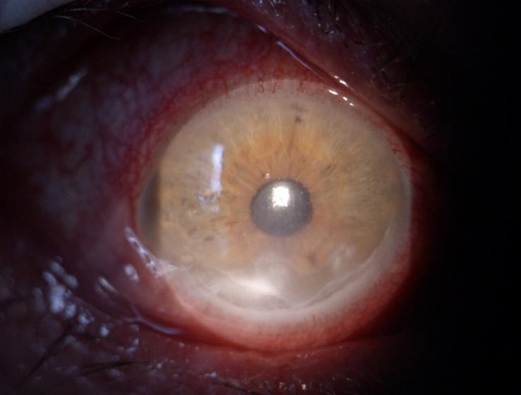
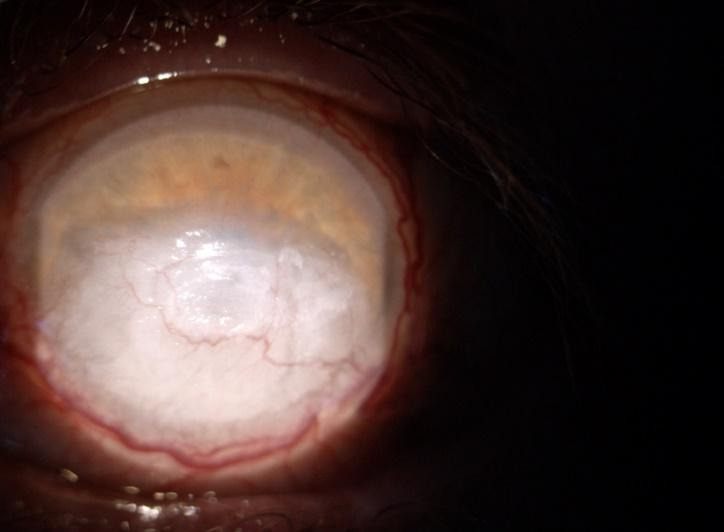
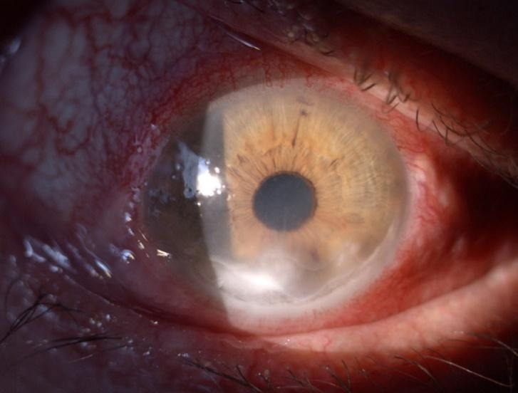
Objective: Our aim was to present a rare case of a middle-aged male patient, diagnosed with Graves' orbitopathy, which had an atypical rapid unilateral onset. Initially, the left eye presented exophthalmos, eyelid retraction, corneal ulceration, and pannus formation with an important vascular component due to corneal exposure. The same symptoms developed in the right eye within a short period of time. Methods: A 52-year-old man presented in our department with bilateral proptosis, decrease in visual acuity, and orbital pain, which developed initially in the left eye seven months before the right eye. Slit lamp examination revealed conjunctival hyperemia, purulent discharge, chemosis and inflammation of the caruncle in both eyes. The fluorescein eye stain test was positive due to corneal ulceration with the presence of cells and flare in anterior chamber in the RE (right eye). The LE (left eye) presented a corneal pannus. We documented the changes using a slit lamp biomicroscope, a fundus camera, orbital ultrasonography, and contrast CT (computer tomography) scans. Discussions: The severe Graves' ophthalmopathy represents a challenge both in active or inactive phase. Medical and surgical therapies should be taken into consideration in order to prevent the complications following corneal perforation or optic neuropathy. Also, ophthalmic, and systemic adverse reactions of systemic steroids used in the treatment of Graves' disease are important in the prognosis of the visual outcome. Conclusions: The management of Graves' ophthalmopathy is multidisciplinary and needs a very good therapy adherence in order to achieve a satisfactory prognosis and quality of life.

 求助内容:
求助内容: 应助结果提醒方式:


