Weixiao Li , Jun Li , Qiang li , Mingzhe Cui , Rutao Xu , Shuiting Zhai , Cheshire Nick , Tianxiao Li , Jiangbo Chen , Wenli Zhao
{"title":"A canine model of aortic arch aneurysm created with autologous pericardium","authors":"Weixiao Li , Jun Li , Qiang li , Mingzhe Cui , Rutao Xu , Shuiting Zhai , Cheshire Nick , Tianxiao Li , Jiangbo Chen , Wenli Zhao","doi":"10.1016/j.jimed.2022.06.005","DOIUrl":null,"url":null,"abstract":"<div><h3>Background</h3><p>To establish a canine model of aortic arch aneurysm that is suitable for research on new devices and techniques applied to the aortic arch.</p></div><div><h3>Materials and methods</h3><p>Fifteen mongrel dogs underwent surgery. The autologous pericardial patch was sewn on the aortotomy site in the anterior wall of the aortic arch. The animals were followed up for 3 months postoperatively. Computed tomography angiography was used to visualize and measure the aneurysm model. Hematoxylin and eosin staining was used to observe the histological characteristics of the aneurysm model. Changes in aneurysm diameter over time were analyzed using analysis of variance.</p></div><div><h3>Results</h3><p>One dog died of hemorrhage during surgery. Fourteen dogs survived the surgical procedure. Two of them died on the first postoperative day because of ruptures at the suturing margin. The diameter of the aneurysm model was twice as large as that of the aortic arch. There was no significant change in the maximum diameter of the aneurysm model during the follow-up period.</p></div><div><h3>Conclusions</h3><p>We established a controllable and stable aortic arch aneurysm model created with an autologous pericardium patch. The aneurysm model can be used to research endoleaks after thoracic endovascular aortic repair and new endovascular techniques can be applied to the aortic arch.</p></div>","PeriodicalId":33533,"journal":{"name":"Journal of Interventional Medicine","volume":"5 3","pages":"Pages 133-137"},"PeriodicalIF":0.0000,"publicationDate":"2022-08-01","publicationTypes":"Journal Article","fieldsOfStudy":null,"isOpenAccess":false,"openAccessPdf":"https://ftp.ncbi.nlm.nih.gov/pub/pmc/oa_pdf/72/bd/main.PMC9617150.pdf","citationCount":"0","resultStr":null,"platform":"Semanticscholar","paperid":null,"PeriodicalName":"Journal of Interventional Medicine","FirstCategoryId":"3","ListUrlMain":"https://www.sciencedirect.com/science/article/pii/S2096360222000394","RegionNum":0,"RegionCategory":null,"ArticlePicture":[],"TitleCN":null,"AbstractTextCN":null,"PMCID":null,"EPubDate":"","PubModel":"","JCR":"Q3","JCRName":"Medicine","Score":null,"Total":0}
引用次数: 0
Abstract
Background
To establish a canine model of aortic arch aneurysm that is suitable for research on new devices and techniques applied to the aortic arch.
Materials and methods
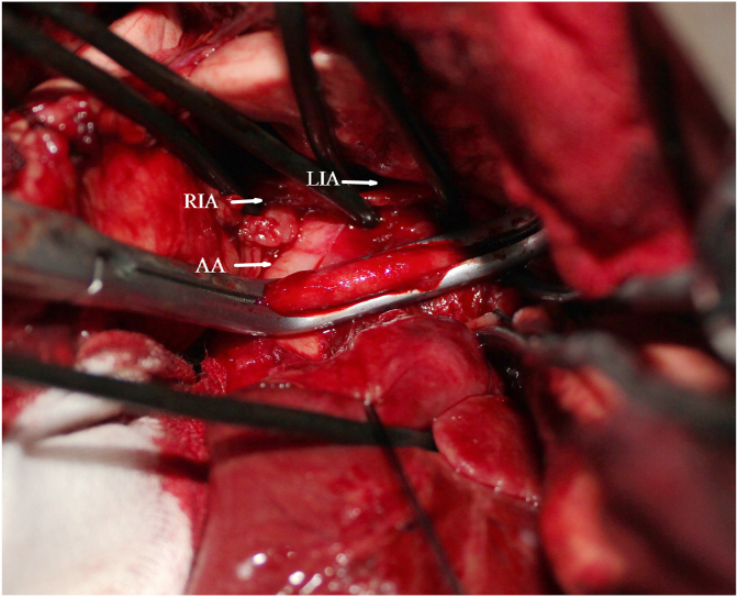
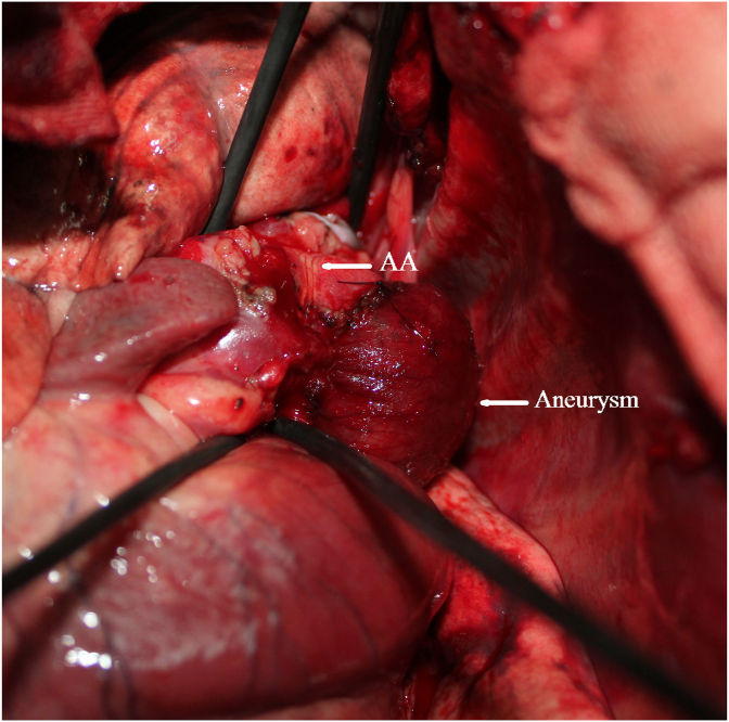
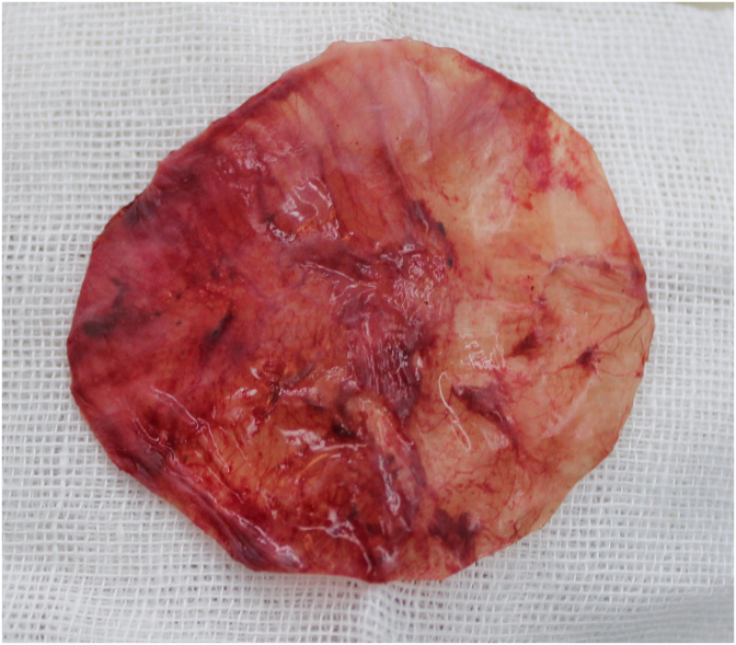
Fifteen mongrel dogs underwent surgery. The autologous pericardial patch was sewn on the aortotomy site in the anterior wall of the aortic arch. The animals were followed up for 3 months postoperatively. Computed tomography angiography was used to visualize and measure the aneurysm model. Hematoxylin and eosin staining was used to observe the histological characteristics of the aneurysm model. Changes in aneurysm diameter over time were analyzed using analysis of variance.
Results
One dog died of hemorrhage during surgery. Fourteen dogs survived the surgical procedure. Two of them died on the first postoperative day because of ruptures at the suturing margin. The diameter of the aneurysm model was twice as large as that of the aortic arch. There was no significant change in the maximum diameter of the aneurysm model during the follow-up period.
Conclusions
We established a controllable and stable aortic arch aneurysm model created with an autologous pericardium patch. The aneurysm model can be used to research endoleaks after thoracic endovascular aortic repair and new endovascular techniques can be applied to the aortic arch.
 求助内容:
求助内容: 应助结果提醒方式:


