hUCMSCs reduce theca interstitial cells apoptosis and restore ovarian function in premature ovarian insufficiency rats through regulating NR4A1-mediated mitochondrial mechanisms.
{"title":"hUCMSCs reduce theca interstitial cells apoptosis and restore ovarian function in premature ovarian insufficiency rats through regulating NR4A1-mediated mitochondrial mechanisms.","authors":"Qianqian Luo, Yu Tang, Zhonglin Jiang, Hongchu Bao, Qiang Fu, Hongqin Zhang","doi":"10.1186/s12958-022-00992-5","DOIUrl":null,"url":null,"abstract":"<p><strong>Background: </strong>Human umbilical cord mesenchymal stem cells (hUCMSCs, retrospectively registered) have a lot of promise for treating theca interstitial cells(TICs) dysfunction in premature ovarian insufficiency (POI). The mechanisms, however, are still unknown.</p><p><strong>Methods: </strong>To examine the therapeutic and find the cause, we used both in vivo cisplatin-induced POI rat model and in vitro TICs model. HUCMSCs were injected into the tail veins of POI rats in an in vivo investigation. Then, using ELISA, HE staining, TUNEL apoptosis test kit, immunohistochemistry and western blot, researchers examined hormonal levels, ovarian morphology, TICs apoptosis, NR4A1 and Cyp17a1 in response to cisplatin treatment and hUCMSCs. TICs were obtained from the ovaries of rats and treated with the cisplatin, hUCMSCs supernatant, and the antagonist of NR4A1--DIM-C-pPhOH. ELISA, immunofluorescence, flow cytometry, JC-1 labeling and western blot analysis were used to detect T levels, Cyp17a1, NR4A1, and the anti-apoptotic protein Bcl-2, as well as pro-apoptotic proteins Bax, caspase-9, caspase-3, and cytochrome C(cytc).</p><p><strong>Results: </strong>We discovered that hUCMSCs restored the ovarian function, particularly TICs function based on measures of Cyp17a1 and T expression. NR4A1 was found in ovarian TICs of each group and NR4A1 expression was lower in the POI rats but higher following hUCMSCs therapy. The apoptosis of TICs generated by cisplatin was reduced after treatment with hUCMSCs. In vitro, NR4A1 was expressed in the nucleus of TICs, and NR4A1 as well as phospho-NR4A1 were decreased, following the apoptosis of TICs was emerged after cisplatin treatment. Interestingly, the localization of NR4A1 was translocated from the nucleus to the cytoplasm due to cisplatin. HUCMSCs were able to boost NR4A1 and phospho-NR4A1 expression while TICs' apoptosis and JC-1 polymorimonomor fluorescence ratios reduced. Furthermore, Bcl-2 expression dropped following cisplatin treatment, whereas Bax, cytc, caspase-9, and caspase-3 expression rose; however, hUCMSCs treatment reduced their expression. In addition, DIM-C-pPhOH had no effect on the NR4A1 expression, but it did increase the expression of apoptosis-related factors such as Bax, cytc, caspase-9, and caspase-3, causing the apoptosis of TICs.</p><p><strong>Conclusions: </strong>These data show that hUCMSCs therapy improves ovarian function in POI rats by inhibiting TICs apoptosis through regulating NR4A1 -mediated mitochondrial mechanisms.</p>","PeriodicalId":520764,"journal":{"name":"Reproductive biology and endocrinology : RB&E","volume":" ","pages":"125"},"PeriodicalIF":0.0000,"publicationDate":"2022-08-19","publicationTypes":"Journal Article","fieldsOfStudy":null,"isOpenAccess":false,"openAccessPdf":"https://www.ncbi.nlm.nih.gov/pmc/articles/PMC9389823/pdf/","citationCount":"4","resultStr":null,"platform":"Semanticscholar","paperid":null,"PeriodicalName":"Reproductive biology and endocrinology : RB&E","FirstCategoryId":"3","ListUrlMain":"https://doi.org/10.1186/s12958-022-00992-5","RegionNum":0,"RegionCategory":null,"ArticlePicture":[],"TitleCN":null,"AbstractTextCN":null,"PMCID":null,"EPubDate":"","PubModel":"","JCR":"","JCRName":"","Score":null,"Total":0}
引用次数: 4
Abstract
Background: Human umbilical cord mesenchymal stem cells (hUCMSCs, retrospectively registered) have a lot of promise for treating theca interstitial cells(TICs) dysfunction in premature ovarian insufficiency (POI). The mechanisms, however, are still unknown.
Methods: To examine the therapeutic and find the cause, we used both in vivo cisplatin-induced POI rat model and in vitro TICs model. HUCMSCs were injected into the tail veins of POI rats in an in vivo investigation. Then, using ELISA, HE staining, TUNEL apoptosis test kit, immunohistochemistry and western blot, researchers examined hormonal levels, ovarian morphology, TICs apoptosis, NR4A1 and Cyp17a1 in response to cisplatin treatment and hUCMSCs. TICs were obtained from the ovaries of rats and treated with the cisplatin, hUCMSCs supernatant, and the antagonist of NR4A1--DIM-C-pPhOH. ELISA, immunofluorescence, flow cytometry, JC-1 labeling and western blot analysis were used to detect T levels, Cyp17a1, NR4A1, and the anti-apoptotic protein Bcl-2, as well as pro-apoptotic proteins Bax, caspase-9, caspase-3, and cytochrome C(cytc).
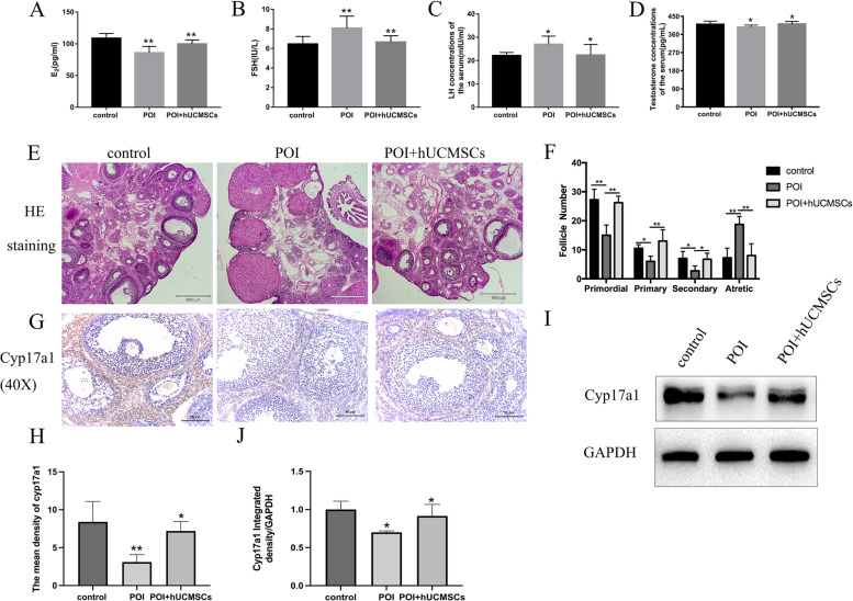
Results: We discovered that hUCMSCs restored the ovarian function, particularly TICs function based on measures of Cyp17a1 and T expression. NR4A1 was found in ovarian TICs of each group and NR4A1 expression was lower in the POI rats but higher following hUCMSCs therapy. The apoptosis of TICs generated by cisplatin was reduced after treatment with hUCMSCs. In vitro, NR4A1 was expressed in the nucleus of TICs, and NR4A1 as well as phospho-NR4A1 were decreased, following the apoptosis of TICs was emerged after cisplatin treatment. Interestingly, the localization of NR4A1 was translocated from the nucleus to the cytoplasm due to cisplatin. HUCMSCs were able to boost NR4A1 and phospho-NR4A1 expression while TICs' apoptosis and JC-1 polymorimonomor fluorescence ratios reduced. Furthermore, Bcl-2 expression dropped following cisplatin treatment, whereas Bax, cytc, caspase-9, and caspase-3 expression rose; however, hUCMSCs treatment reduced their expression. In addition, DIM-C-pPhOH had no effect on the NR4A1 expression, but it did increase the expression of apoptosis-related factors such as Bax, cytc, caspase-9, and caspase-3, causing the apoptosis of TICs.
Conclusions: These data show that hUCMSCs therapy improves ovarian function in POI rats by inhibiting TICs apoptosis through regulating NR4A1 -mediated mitochondrial mechanisms.

 求助内容:
求助内容: 应助结果提醒方式:


