Cortical spreading depression can be triggered by sensory stimulation in primed wild type mouse brain: a mechanistic insight to migraine aura generation.
{"title":"Cortical spreading depression can be triggered by sensory stimulation in primed wild type mouse brain: a mechanistic insight to migraine aura generation.","authors":"Sahin Hanalioglu, Aslihan Taskiran-Sag, Hulya Karatas, Buket Donmez-Demir, Sinem Yilmaz-Ozcan, Emine Eren-Kocak, Yasemin Gursoy-Ozdemir, Turgay Dalkara","doi":"10.1186/s10194-022-01474-0","DOIUrl":null,"url":null,"abstract":"<p><strong>Background: </strong>Unlike the spontaneously appearing aura in migraineurs, experimentally, cortical spreading depression (CSD), the neurophysiological correlate of aura is induced by non-physiological stimuli. Consequently, neural mechanisms involved in spontaneous CSD generation, which may provide insight into how migraine starts in an otherwise healthy brain, remain largely unclear. We hypothesized that CSD can be physiologically induced by sensory stimulation in primed mouse brain.</p><p><strong>Methods: </strong>Cortex was made susceptible to CSD with partial inhibition of Na<sup>+</sup>/K<sup>+</sup>-ATPase by epidural application of a low concentration of Na<sup>+</sup>/K<sup>+</sup>-ATPase blocker ouabain, allowing longer than 30-min intervals between CSDs or by knocking-down α2 subunit of Na<sup>+</sup>/K<sup>+</sup>-ATPase, which is crucial for K<sup>+</sup> and glutamate re-uptake, with shRNA. Stimulation-triggered CSDs and extracellular K<sup>+</sup> changes were monitored in vivo electrophysiologically and a K<sup>+</sup>-sensitive fluoroprobe (IPG-4), respectively.</p><p><strong>Results: </strong>After priming with ouabain, photic stimulation significantly increased the CSD incidence compared with non-stimulated animals (44.0 vs. 4.9%, p < 0.001). Whisker stimulation also significantly increased the CSD incidence, albeit less effectively (14.9 vs. 2.4%, p = 0.02). Knocking-down Na<sup>+</sup>/K<sup>+</sup>-ATPase (50% decrease in mRNA) lowered the CSD threshold in all mice tested with KCl but triggered CSDs in 14.3% and 16.7% of mice with photic and whisker stimulation, respectively. Confirming Na<sup>+</sup>/K<sup>+</sup>-ATPase hypofunction, extracellular K<sup>+</sup> significantly rose during sensory stimulation after ouabain or shRNA treatment unlike controls. In line with the higher CSD susceptibility observed, K<sup>+</sup> rise was more prominent after ouabain. To gain insight to preventive mechanisms reducing the probability of stimulus-evoked CSDs, we applied an A1-receptor antagonist (DPCPX) to the occipital cortex, because adenosine formed during stimulation from ATP can reduce CSD susceptibility. DPCPX induced spontaneous CSDs but only small-DC shifts along with suppression of EEG spikes during photic stimulation, suggesting that the inhibition co-activated with sensory stimulation could limit CSD ignition when K<sup>+</sup> uptake was not sufficiently suppressed as with ouabain.</p><p><strong>Conclusions: </strong>Normal brain is well protected against CSD generation. For CSD to be ignited under physiological conditions, priming and predisposing factors are required as seen in migraine patients. Intense sensory stimulation has potential to trigger CSD when co-existing conditions bring extracellular K<sup>+</sup> and glutamate concentrations over CSD-ignition threshold and stimulation-evoked inhibitory mechanisms are overcome.</p>","PeriodicalId":501630,"journal":{"name":"The Journal of Headache and Pain","volume":" ","pages":"107"},"PeriodicalIF":0.0000,"publicationDate":"2022-08-19","publicationTypes":"Journal Article","fieldsOfStudy":null,"isOpenAccess":false,"openAccessPdf":"https://www.ncbi.nlm.nih.gov/pmc/articles/PMC9392331/pdf/","citationCount":"5","resultStr":null,"platform":"Semanticscholar","paperid":null,"PeriodicalName":"The Journal of Headache and Pain","FirstCategoryId":"3","ListUrlMain":"https://doi.org/10.1186/s10194-022-01474-0","RegionNum":0,"RegionCategory":null,"ArticlePicture":[],"TitleCN":null,"AbstractTextCN":null,"PMCID":null,"EPubDate":"","PubModel":"","JCR":"","JCRName":"","Score":null,"Total":0}
引用次数: 5
Abstract
Background: Unlike the spontaneously appearing aura in migraineurs, experimentally, cortical spreading depression (CSD), the neurophysiological correlate of aura is induced by non-physiological stimuli. Consequently, neural mechanisms involved in spontaneous CSD generation, which may provide insight into how migraine starts in an otherwise healthy brain, remain largely unclear. We hypothesized that CSD can be physiologically induced by sensory stimulation in primed mouse brain.
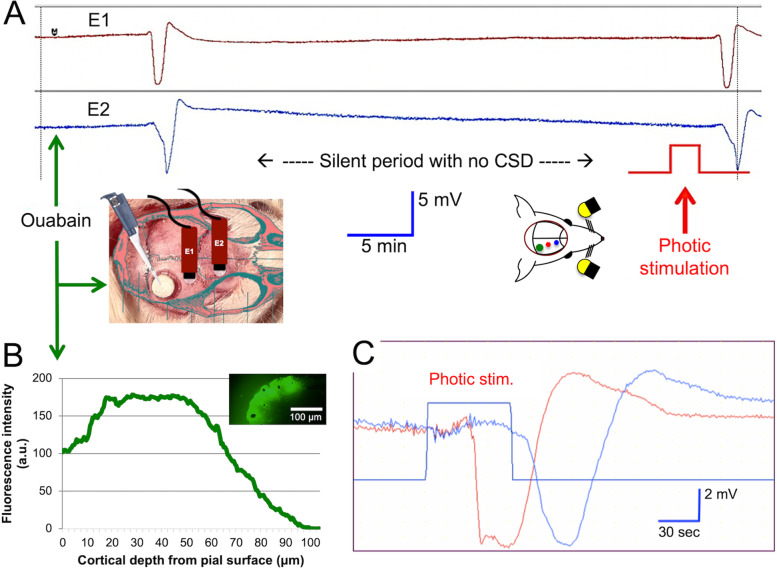
Methods: Cortex was made susceptible to CSD with partial inhibition of Na+/K+-ATPase by epidural application of a low concentration of Na+/K+-ATPase blocker ouabain, allowing longer than 30-min intervals between CSDs or by knocking-down α2 subunit of Na+/K+-ATPase, which is crucial for K+ and glutamate re-uptake, with shRNA. Stimulation-triggered CSDs and extracellular K+ changes were monitored in vivo electrophysiologically and a K+-sensitive fluoroprobe (IPG-4), respectively.
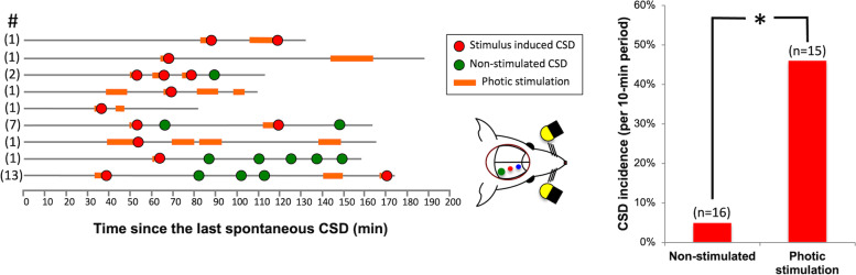
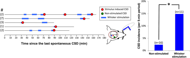
Results: After priming with ouabain, photic stimulation significantly increased the CSD incidence compared with non-stimulated animals (44.0 vs. 4.9%, p < 0.001). Whisker stimulation also significantly increased the CSD incidence, albeit less effectively (14.9 vs. 2.4%, p = 0.02). Knocking-down Na+/K+-ATPase (50% decrease in mRNA) lowered the CSD threshold in all mice tested with KCl but triggered CSDs in 14.3% and 16.7% of mice with photic and whisker stimulation, respectively. Confirming Na+/K+-ATPase hypofunction, extracellular K+ significantly rose during sensory stimulation after ouabain or shRNA treatment unlike controls. In line with the higher CSD susceptibility observed, K+ rise was more prominent after ouabain. To gain insight to preventive mechanisms reducing the probability of stimulus-evoked CSDs, we applied an A1-receptor antagonist (DPCPX) to the occipital cortex, because adenosine formed during stimulation from ATP can reduce CSD susceptibility. DPCPX induced spontaneous CSDs but only small-DC shifts along with suppression of EEG spikes during photic stimulation, suggesting that the inhibition co-activated with sensory stimulation could limit CSD ignition when K+ uptake was not sufficiently suppressed as with ouabain.
Conclusions: Normal brain is well protected against CSD generation. For CSD to be ignited under physiological conditions, priming and predisposing factors are required as seen in migraine patients. Intense sensory stimulation has potential to trigger CSD when co-existing conditions bring extracellular K+ and glutamate concentrations over CSD-ignition threshold and stimulation-evoked inhibitory mechanisms are overcome.
背景:不同于偏头痛自发出现的先兆,实验表明,皮层扩张性抑制(CSD),先兆的神经生理关联是由非生理性刺激引起的。因此,参与自发性CSD产生的神经机制,可能为了解偏头痛如何在健康的大脑中开始提供见解,在很大程度上仍然不清楚。我们假设CSD可以通过刺激小鼠大脑的感觉来生理诱导。方法:通过硬膜外应用低浓度的Na+/K+- atp酶阻滞剂瓦巴因,使CSD间隔超过30分钟,或通过shRNA敲低Na+/K+- atp酶的α2亚基(对K+和谷氨酸的再摄取至关重要),使皮质对CSD敏感。刺激触发的CSDs和细胞外K+变化分别在体内电生理和K+敏感氟探针(IPG-4)中监测。结果:与未刺激的小鼠相比,光刺激后的CSD发生率显著增加(44.0% vs. 4.9%), p +/K+- atp酶(mRNA减少50%)降低了所有KCl小鼠的CSD阈值,但光刺激和须刺激小鼠的CSD发生率分别为14.3%和16.7%。证实Na+/K+- atp酶功能低下,细胞外K+在沃阿因或shRNA治疗后与对照组不同,在感觉刺激期间显著升高。与观察到的较高的CSD敏感性一致,沃巴因后K+升高更为明显。为了深入了解降低刺激诱发CSD可能性的预防机制,我们将a1受体拮抗剂(DPCPX)应用于枕皮质,因为ATP在刺激过程中形成的腺苷可以降低CSD的易感性。DPCPX诱导自发性CSD,但在光刺激下仅发生小的dc偏移,同时抑制脑电图峰值,这表明当K+摄取不像瓦巴因那样被充分抑制时,与感觉刺激共同激活的抑制可以限制CSD的点燃。结论:正常脑对CSD的产生具有良好的保护作用。如偏头痛患者所见,CSD在生理条件下被点燃,需要启动和诱发因素。当共存条件使细胞外K+和谷氨酸浓度超过CSD点燃阈值并克服刺激诱发的抑制机制时,强烈的感觉刺激有可能触发CSD。

 求助内容:
求助内容: 应助结果提醒方式:


