{"title":"Intraperitoneal bleeding from the right gastroepiploic artery by endoscopic ultrasonography: a case report.","authors":"Koji Takahashi, Hiroshi Ohyama, Rintaro Mikata, Hiroki Nagashima, Izumi Ohno, Yuichi Takiguchi, Naoya Kato","doi":"10.2185/jrm.2022-002","DOIUrl":null,"url":null,"abstract":"<p><p><b>Objective:</b> To describe the case of a patient with intraperitoneal bleeding from the gastroepiploic artery by endoscopic ultrasound who was successfully treated with transcatheter arterial coil embolization. <b>Patient and Methods:</b> An 87-year-old man was referred to our hospital for examination of a gallbladder tumor. Endoscopic ultrasonography was performed using an oblique-view echoendoscope. After the endoscopic ultrasound, the patient went into shock. Computed tomography revealed a huge intraperitoneal hematoma and an aneurysm in the right gastroepiploic artery that were not seen on previous computed tomography images. Thus, urgent catheter angiography was performed, which showed a pseudoaneurysm of the right gastroepiploic artery and extravasation of the contrast medium from the pseudoaneurysm. <b>Results:</b> Transcatheter arterial coil embolization was subsequently performed, and the bleeding stopped. Thereafter, his hemodynamics stabilized and his general condition improved. The patient was discharged 22 days post-treatment with an uneventful course. <b>Conclusion:</b> Observation-only endoscopic ultrasound without invasive procedures can cause intraperitoneal bleeding due to a ruptured splanchnic artery. Thus, endoscopic ultrasonography should be performed more carefully in elderly patients.</p>","PeriodicalId":73939,"journal":{"name":"Journal of rural medicine : JRM","volume":"17 3","pages":"184-188"},"PeriodicalIF":0.0000,"publicationDate":"2022-07-01","publicationTypes":"Journal Article","fieldsOfStudy":null,"isOpenAccess":false,"openAccessPdf":"https://ftp.ncbi.nlm.nih.gov/pub/pmc/oa_pdf/7f/b2/jrm-17-184.PMC9263947.pdf","citationCount":"0","resultStr":null,"platform":"Semanticscholar","paperid":null,"PeriodicalName":"Journal of rural medicine : JRM","FirstCategoryId":"1085","ListUrlMain":"https://doi.org/10.2185/jrm.2022-002","RegionNum":0,"RegionCategory":null,"ArticlePicture":[],"TitleCN":null,"AbstractTextCN":null,"PMCID":null,"EPubDate":"","PubModel":"","JCR":"","JCRName":"","Score":null,"Total":0}
引用次数: 0
Abstract
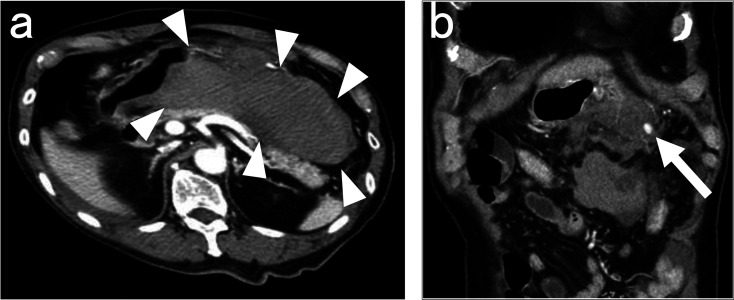
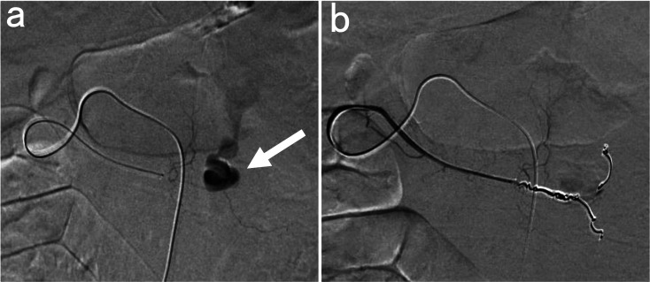
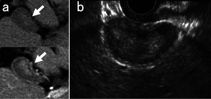
Objective: To describe the case of a patient with intraperitoneal bleeding from the gastroepiploic artery by endoscopic ultrasound who was successfully treated with transcatheter arterial coil embolization. Patient and Methods: An 87-year-old man was referred to our hospital for examination of a gallbladder tumor. Endoscopic ultrasonography was performed using an oblique-view echoendoscope. After the endoscopic ultrasound, the patient went into shock. Computed tomography revealed a huge intraperitoneal hematoma and an aneurysm in the right gastroepiploic artery that were not seen on previous computed tomography images. Thus, urgent catheter angiography was performed, which showed a pseudoaneurysm of the right gastroepiploic artery and extravasation of the contrast medium from the pseudoaneurysm. Results: Transcatheter arterial coil embolization was subsequently performed, and the bleeding stopped. Thereafter, his hemodynamics stabilized and his general condition improved. The patient was discharged 22 days post-treatment with an uneventful course. Conclusion: Observation-only endoscopic ultrasound without invasive procedures can cause intraperitoneal bleeding due to a ruptured splanchnic artery. Thus, endoscopic ultrasonography should be performed more carefully in elderly patients.

 求助内容:
求助内容: 应助结果提醒方式:


