Laura Cacciaguerra, Damiano Mistri, Paola Valsasina, Vittorio Martinelli, Massimo Filippi, Maria A Rocca
{"title":"Time-varying connectivity of the precuneus and its association with cognition and depressive symptoms in neuromyelitis optica: A pilot MRI study.","authors":"Laura Cacciaguerra, Damiano Mistri, Paola Valsasina, Vittorio Martinelli, Massimo Filippi, Maria A Rocca","doi":"10.1177/13524585221107125","DOIUrl":null,"url":null,"abstract":"<p><strong>Background: </strong>The precuneus is involved in cognition and depression; static functional connectivity (SFC) abnormalities of this region have been observed in neuromyelitis optica spectrum disorders (NMOSD). Time-varying functional connectivity (TVC) underpins dynamic variations of brain connectivity.</p><p><strong>Objective: </strong>The aim of this study was to explore precuneus SFC and TVC in NMOSD patients and their associations with neuropsychological features.</p><p><strong>Methods: </strong>This retrospective study includes 27 NMOSD patients and 30 matched healthy controls undergoing resting state functional magnetic resonance imaging (MRI) and a neuropsychological evaluation of cognitive performance and depressive symptoms. A sliding-window correlation analysis using bilateral precuneus as seed region assessed TVC, which was quantified by the standard deviation of connectivity across windows. Mean connectivity indicated SFC.</p><p><strong>Results: </strong>Compared to controls, patients had reduced SFC between precuneus, temporal lobe, putamen and cerebellum, and reduced TVC between precuneus and prefronto-parietal-temporo-occipital cortices and caudate. Patients also had increased intra-precuneal TVC and increased TVC between the precuneus and the temporal cortex. More severe depressive symptoms correlated with increased TVC between the precuneus and the temporal lobe; worse cognitive performance mainly correlated with higher TVC between the precuneus and the parietal lobe.</p><p><strong>Conclusion: </strong>TVC rather than SFC of the precuneus correlates with NMOSD neuropsychological features; different TVC abnormalities underlie depressive symptoms and cognitive impairment.</p>","PeriodicalId":520714,"journal":{"name":"Multiple sclerosis (Houndmills, Basingstoke, England)","volume":" ","pages":"2057-2069"},"PeriodicalIF":0.0000,"publicationDate":"2022-11-01","publicationTypes":"Journal Article","fieldsOfStudy":null,"isOpenAccess":false,"openAccessPdf":"https://ftp.ncbi.nlm.nih.gov/pub/pmc/oa_pdf/a1/f8/10.1177_13524585221107125.PMC9574904.pdf","citationCount":"2","resultStr":null,"platform":"Semanticscholar","paperid":null,"PeriodicalName":"Multiple sclerosis (Houndmills, Basingstoke, England)","FirstCategoryId":"3","ListUrlMain":"https://doi.org/10.1177/13524585221107125","RegionNum":0,"RegionCategory":null,"ArticlePicture":[],"TitleCN":null,"AbstractTextCN":null,"PMCID":null,"EPubDate":"2022/7/7 0:00:00","PubModel":"Epub","JCR":"","JCRName":"","Score":null,"Total":0}
引用次数: 2
Abstract
Background: The precuneus is involved in cognition and depression; static functional connectivity (SFC) abnormalities of this region have been observed in neuromyelitis optica spectrum disorders (NMOSD). Time-varying functional connectivity (TVC) underpins dynamic variations of brain connectivity.
Objective: The aim of this study was to explore precuneus SFC and TVC in NMOSD patients and their associations with neuropsychological features.
Methods: This retrospective study includes 27 NMOSD patients and 30 matched healthy controls undergoing resting state functional magnetic resonance imaging (MRI) and a neuropsychological evaluation of cognitive performance and depressive symptoms. A sliding-window correlation analysis using bilateral precuneus as seed region assessed TVC, which was quantified by the standard deviation of connectivity across windows. Mean connectivity indicated SFC.
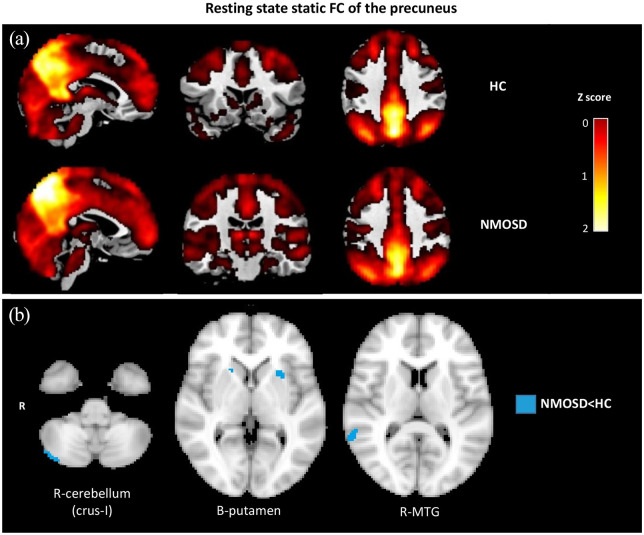
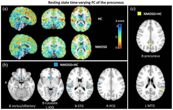
Results: Compared to controls, patients had reduced SFC between precuneus, temporal lobe, putamen and cerebellum, and reduced TVC between precuneus and prefronto-parietal-temporo-occipital cortices and caudate. Patients also had increased intra-precuneal TVC and increased TVC between the precuneus and the temporal cortex. More severe depressive symptoms correlated with increased TVC between the precuneus and the temporal lobe; worse cognitive performance mainly correlated with higher TVC between the precuneus and the parietal lobe.
Conclusion: TVC rather than SFC of the precuneus correlates with NMOSD neuropsychological features; different TVC abnormalities underlie depressive symptoms and cognitive impairment.

 求助内容:
求助内容: 应助结果提醒方式:


