{"title":"Identification of heterogeneous subsets of aortic interleukin-17A-expressing CD4<sup>+</sup> T cells in atherosclerotic mice.","authors":"Guizhen Lin, Lei Zhang, Zheng Yan, Wei Jiang, Beibei Wu, Dongsheng Li, Xiaofang Xiong","doi":"10.1177/03946320221117933","DOIUrl":null,"url":null,"abstract":"<p><strong>Objectives: </strong>T helper 17 (Th17) cells are involved in the inflammatory response of atherosclerosis. However, their heterogeneity in the atherosclerotic aorta remains elusive. This study was designed to identify aortic Th17 subsets.</p><p><strong>Methods: </strong>The surface markers and transcription factors of aortic interleukin-17A (IL-17A)-expressing T cells were determined by flow cytometry in an ApoE-deficient mouse atherosclerotic model. Viable aortic IL-17A-expressing T cell subsets were isolated by flow cytometry on the basis of surface markers, followed by characterizing their transcription factors by either flow cytometry or real-time RT-PCR. The effect of aortic IL-17A-expressing T cell subsets on aortic endothelial cells was determined in vitro.</p><p><strong>Results: </strong>C-X-C Motif Chemokine Receptor 3 (CXCR3), interleukin-17 receptor E (IL-17RE), CD200, and C-C Motif Chemokine Receptor 4 (CCR4) marked three subsets of aortic IL-17A-expressing T cells: CXCR3<sup>+</sup>IL-17RE<sup>low</sup>CD200<sup>+</sup>CCR4<sup>-</sup> T cells expressing T-box protein expressed in T cells (T-bet) and interferon-gamma (IFN-γ), CXCR3<sup>+</sup>IL-17RE<sup>low</sup>CD200<sup>+</sup>CCR4<sup>+</sup> T cells expressing T-bet but fewer IFN-γ, and CXCR3<sup>-</sup>IL-17RE<sup>high</sup>CD200<sup>+</sup>CCR4<sup>+</sup> T cells expressing very low T-bet and no IFN-γ. Based on these markers, viable aortic Th17 cells, Th17.1 cells, and transitional Th17.1 cells were identified. Both Th17.1 cells and transitional Th17.1 cells were more proliferative than Th17 cells. Compared with Th17 cells, Th17.1 cells plus transitional Th17.1 cells induced higher expression of C-X-C motif chemokine ligand 1 (CXCL1), C-C motif chemokine ligand 2 (CCL2), C-X-C motif chemokine 5 (CXCL5), and granulocyte-macrophage colony-stimulating factor (GM-CSF) in aortic endothelial cells.</p><p><strong>Conclusion: </strong>IL-17A-expressing CD4<sup>+</sup> T cells were heterogeneous in atherosclerotic aortas.</p>","PeriodicalId":14046,"journal":{"name":"International Journal of Immunopathology and Pharmacology","volume":" ","pages":"3946320221117933"},"PeriodicalIF":2.6000,"publicationDate":"2022-01-01","publicationTypes":"Journal Article","fieldsOfStudy":null,"isOpenAccess":false,"openAccessPdf":"https://ftp.ncbi.nlm.nih.gov/pub/pmc/oa_pdf/d6/7d/10.1177_03946320221117933.PMC9364180.pdf","citationCount":"0","resultStr":null,"platform":"Semanticscholar","paperid":null,"PeriodicalName":"International Journal of Immunopathology and Pharmacology","FirstCategoryId":"3","ListUrlMain":"https://doi.org/10.1177/03946320221117933","RegionNum":3,"RegionCategory":"医学","ArticlePicture":[],"TitleCN":null,"AbstractTextCN":null,"PMCID":null,"EPubDate":"","PubModel":"","JCR":"Q3","JCRName":"IMMUNOLOGY","Score":null,"Total":0}
引用次数: 0
Abstract
Objectives: T helper 17 (Th17) cells are involved in the inflammatory response of atherosclerosis. However, their heterogeneity in the atherosclerotic aorta remains elusive. This study was designed to identify aortic Th17 subsets.
Methods: The surface markers and transcription factors of aortic interleukin-17A (IL-17A)-expressing T cells were determined by flow cytometry in an ApoE-deficient mouse atherosclerotic model. Viable aortic IL-17A-expressing T cell subsets were isolated by flow cytometry on the basis of surface markers, followed by characterizing their transcription factors by either flow cytometry or real-time RT-PCR. The effect of aortic IL-17A-expressing T cell subsets on aortic endothelial cells was determined in vitro.
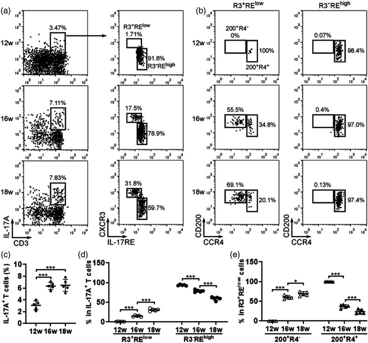
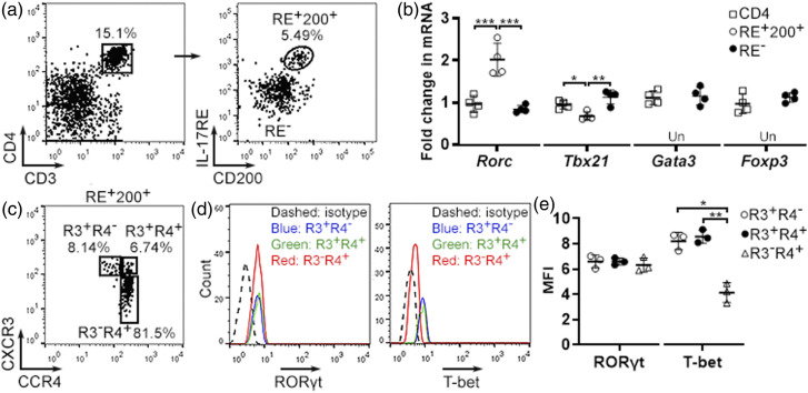
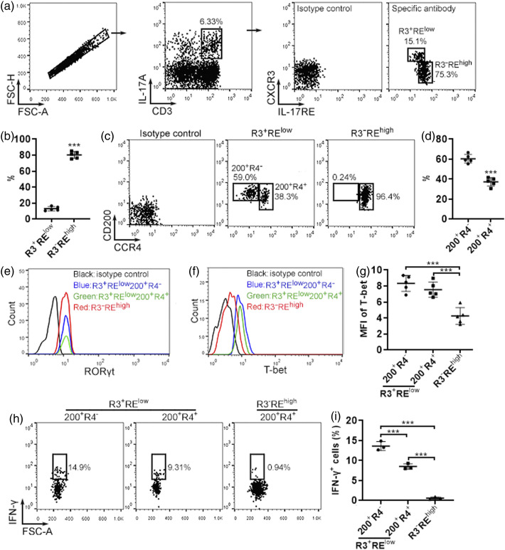
Results: C-X-C Motif Chemokine Receptor 3 (CXCR3), interleukin-17 receptor E (IL-17RE), CD200, and C-C Motif Chemokine Receptor 4 (CCR4) marked three subsets of aortic IL-17A-expressing T cells: CXCR3+IL-17RElowCD200+CCR4- T cells expressing T-box protein expressed in T cells (T-bet) and interferon-gamma (IFN-γ), CXCR3+IL-17RElowCD200+CCR4+ T cells expressing T-bet but fewer IFN-γ, and CXCR3-IL-17REhighCD200+CCR4+ T cells expressing very low T-bet and no IFN-γ. Based on these markers, viable aortic Th17 cells, Th17.1 cells, and transitional Th17.1 cells were identified. Both Th17.1 cells and transitional Th17.1 cells were more proliferative than Th17 cells. Compared with Th17 cells, Th17.1 cells plus transitional Th17.1 cells induced higher expression of C-X-C motif chemokine ligand 1 (CXCL1), C-C motif chemokine ligand 2 (CCL2), C-X-C motif chemokine 5 (CXCL5), and granulocyte-macrophage colony-stimulating factor (GM-CSF) in aortic endothelial cells.
Conclusion: IL-17A-expressing CD4+ T cells were heterogeneous in atherosclerotic aortas.
期刊介绍:
International Journal of Immunopathology and Pharmacology is an Open Access peer-reviewed journal publishing original papers describing research in the fields of immunology, pathology and pharmacology. The intention is that the journal should reflect both the experimental and clinical aspects of immunology as well as advances in the understanding of the pathology and pharmacology of the immune system.
 求助内容:
求助内容: 应助结果提醒方式:


