Magdalena Lerch, Kathrin Schanda, Eliott Lafon, Reinhard Würzner, Sara Mariotto, Alessandro Dinoto, Eva Maria Wendel, Christian Lechner, Harald Hegen, Kevin Rostásy, Thomas Berger, Doris Wilflingseder, Romana Höftberger, Markus Reindl
{"title":"More Efficient Complement Activation by Anti-Aquaporin-4 Compared With Anti-Myelin Oligodendrocyte Glycoprotein Antibodies.","authors":"Magdalena Lerch, Kathrin Schanda, Eliott Lafon, Reinhard Würzner, Sara Mariotto, Alessandro Dinoto, Eva Maria Wendel, Christian Lechner, Harald Hegen, Kevin Rostásy, Thomas Berger, Doris Wilflingseder, Romana Höftberger, Markus Reindl","doi":"10.1212/NXI.0000000000200059","DOIUrl":null,"url":null,"abstract":"<p><strong>Background and objectives: </strong>The objective was to study complement-mediated cytotoxicity induced by immunoglobulin G (IgG) anti-aquaporin-4 antibodies (AQP4-IgG) and anti-myelin oligodendrocyte glycoprotein antibodies (MOG-IgG) in human serum samples from patients suffering from the rare demyelinating diseases of the CNS neuromyelitis optica spectrum disorder (NMOSD) and MOG-IgG-associated disease (MOGAD).</p><p><strong>Methods: </strong>A cell-based assay with HEK293A cells expressing different MOG isoforms (MOGα<sub>1-3</sub>β<sub>1-3</sub>) or AQP4-M23 was used. Cells were incubated with human MOG-IgG or AQP4-IgG-positive serum samples together with active or heat-inactivated human complement, and complement-dependent cytotoxicity (CDC) was measured with a lactate dehydrogenase assay. To further quantify antibody-mediated cell damage, formation of the terminal complement complex (TCC) was analyzed by flow cytometry. In addition, immunocytochemistry of the TCC and complement component 3 (C3) was performed.</p><p><strong>Results: </strong>AQP4-IgG-positive serum samples induced higher CDC and TCC levels than MOG-IgG-positive sera. Notably, both showed a correlation between antibody titers and CDC and also between titers and TCC levels. In addition, all 6 MOG isoforms tested (MOGα<sub>1-3</sub>β<sub>1-3</sub>) could induce at least some CDC; however, the strongest MOG-IgG-induced CDC levels were found on MOGα<sub>1</sub>, MOGα<sub>3</sub>, and MOGβ<sub>1</sub>. Different MOG-IgG binding patterns regarding recognition of different MOG isoforms were investigated, and it was found that MOG-IgG recognizing all 6 isoforms again induced highest CDC levels on MOGα<sub>1</sub> and MOGβ<sub>1</sub>. Furthermore, surface staining of TCC and C3 revealed positive staining on all 6 MOG isoforms tested, as well as on AQP4-M23.</p><p><strong>Discussion: </strong>Both MOG-IgG and AQP4-IgG are able to induce CDC in a titer-dependent manner. However, AQP4-IgG showed markedly higher levels of CDC compared with MOG in vitro on target cells. This further highlights the role of complement in AQP4-IgG-mediated disease and diminishes the importance of complement activation in MOG-IgG-mediated autoimmune disease.</p>","PeriodicalId":520720,"journal":{"name":"Neurology(R) neuroimmunology & neuroinflammation","volume":" ","pages":""},"PeriodicalIF":7.5000,"publicationDate":"2022-11-22","publicationTypes":"Journal Article","fieldsOfStudy":null,"isOpenAccess":false,"openAccessPdf":"https://www.ncbi.nlm.nih.gov/pmc/articles/PMC9682624/pdf/","citationCount":"0","resultStr":null,"platform":"Semanticscholar","paperid":null,"PeriodicalName":"Neurology(R) neuroimmunology & neuroinflammation","FirstCategoryId":"1085","ListUrlMain":"https://doi.org/10.1212/NXI.0000000000200059","RegionNum":0,"RegionCategory":null,"ArticlePicture":[],"TitleCN":null,"AbstractTextCN":null,"PMCID":null,"EPubDate":"2023/1/1 0:00:00","PubModel":"Print","JCR":"","JCRName":"","Score":null,"Total":0}
引用次数: 0
Abstract
Background and objectives: The objective was to study complement-mediated cytotoxicity induced by immunoglobulin G (IgG) anti-aquaporin-4 antibodies (AQP4-IgG) and anti-myelin oligodendrocyte glycoprotein antibodies (MOG-IgG) in human serum samples from patients suffering from the rare demyelinating diseases of the CNS neuromyelitis optica spectrum disorder (NMOSD) and MOG-IgG-associated disease (MOGAD).
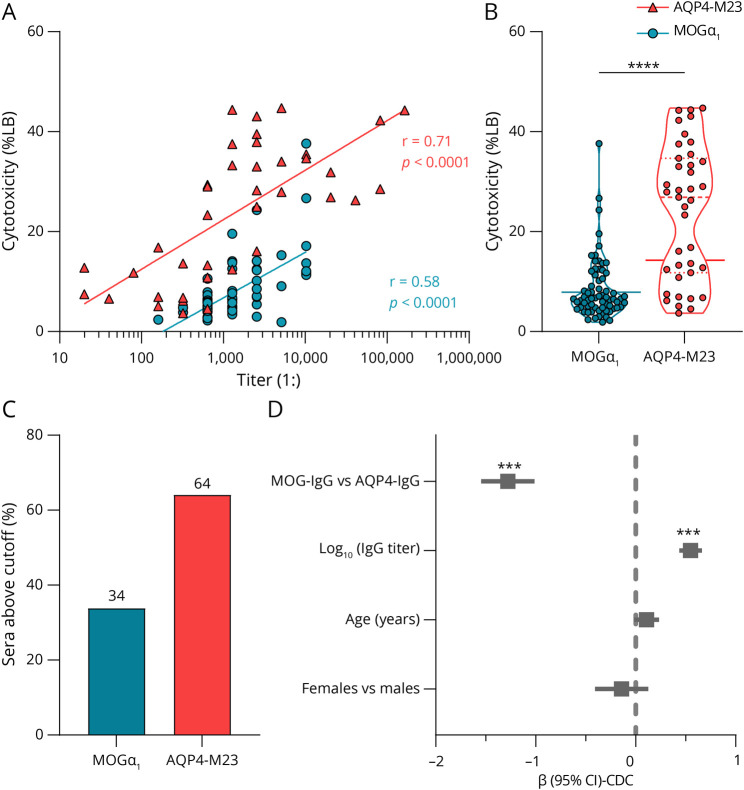
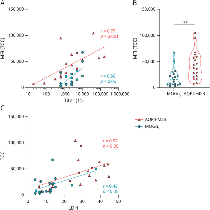
Methods: A cell-based assay with HEK293A cells expressing different MOG isoforms (MOGα1-3β1-3) or AQP4-M23 was used. Cells were incubated with human MOG-IgG or AQP4-IgG-positive serum samples together with active or heat-inactivated human complement, and complement-dependent cytotoxicity (CDC) was measured with a lactate dehydrogenase assay. To further quantify antibody-mediated cell damage, formation of the terminal complement complex (TCC) was analyzed by flow cytometry. In addition, immunocytochemistry of the TCC and complement component 3 (C3) was performed.
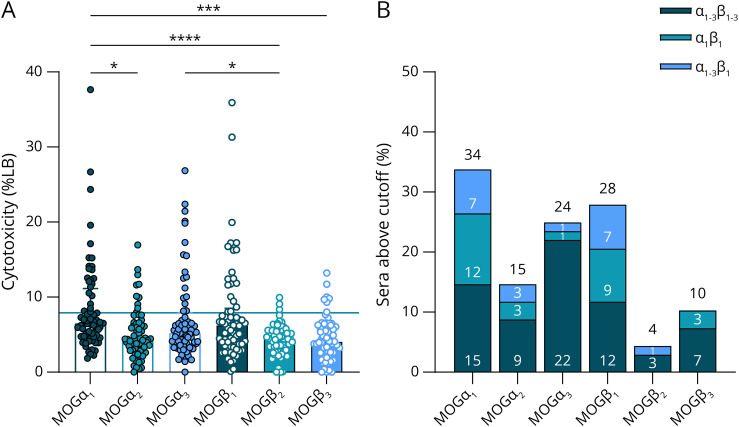
Results: AQP4-IgG-positive serum samples induced higher CDC and TCC levels than MOG-IgG-positive sera. Notably, both showed a correlation between antibody titers and CDC and also between titers and TCC levels. In addition, all 6 MOG isoforms tested (MOGα1-3β1-3) could induce at least some CDC; however, the strongest MOG-IgG-induced CDC levels were found on MOGα1, MOGα3, and MOGβ1. Different MOG-IgG binding patterns regarding recognition of different MOG isoforms were investigated, and it was found that MOG-IgG recognizing all 6 isoforms again induced highest CDC levels on MOGα1 and MOGβ1. Furthermore, surface staining of TCC and C3 revealed positive staining on all 6 MOG isoforms tested, as well as on AQP4-M23.
Discussion: Both MOG-IgG and AQP4-IgG are able to induce CDC in a titer-dependent manner. However, AQP4-IgG showed markedly higher levels of CDC compared with MOG in vitro on target cells. This further highlights the role of complement in AQP4-IgG-mediated disease and diminishes the importance of complement activation in MOG-IgG-mediated autoimmune disease.

 求助内容:
求助内容: 应助结果提醒方式:


