{"title":"Brachialis Muscle Rupture in a Pediatric Patient Followed Up by Ultrasound Examinations: A Rare Case Report.","authors":"Akihiro Yamaji, Masafumi Uesugi, Hiroshi Kamada, Harumitsu Ichimura, Masashi Yamazaki","doi":"10.1155/2022/3391350","DOIUrl":null,"url":null,"abstract":"<p><p>Isolated brachial muscle injuries are relatively rare injuries and reportedly occur during forced elbow extension. Though commonly conservative treatment approach is adopted, the treatment criteria remain unclear. Here, we report the case of a patient who experienced functional recovery after conservative treatment for an isolated brachial muscle injury. The patient was an 8-year-old boy whose chief complaint was left elbow pain. The injury occurred when the patient fell while playing on gymnastics bars and bruised the palmar side of his left elbow on the bar. Owing to the pain in the left elbow, the patient came to our institution. There were no clear signs of deformities or swelling in the left elbow and no obvious tenderness. X-ray and computed tomography (CT) imaging examinations revealed no signs of a fracture or dislocation, and the patient was diagnosed with left brachialis muscle rupture based on magnetic resonance imaging (MRI). Although the brachialis muscle was complete ruptured, a healing tendency was seen on body surface ultrasound examinations over time, and the patient was treated conservatively. After 3 weeks of cast immobilization, the patient underwent range of motion exercises. Two months after the injury, there were no issues with elbow joint function in daily life activities and no limitations in range of motion. Here, MRI was used to diagnose brachialis muscle rupture, and ultrasound examinations were utilized to make treatment decisions.</p>","PeriodicalId":30287,"journal":{"name":"Case Reports in Orthopedics","volume":" ","pages":"3391350"},"PeriodicalIF":0.6000,"publicationDate":"2022-06-23","publicationTypes":"Journal Article","fieldsOfStudy":null,"isOpenAccess":false,"openAccessPdf":"https://www.ncbi.nlm.nih.gov/pmc/articles/PMC9246635/pdf/","citationCount":"0","resultStr":null,"platform":"Semanticscholar","paperid":null,"PeriodicalName":"Case Reports in Orthopedics","FirstCategoryId":"1085","ListUrlMain":"https://doi.org/10.1155/2022/3391350","RegionNum":0,"RegionCategory":null,"ArticlePicture":[],"TitleCN":null,"AbstractTextCN":null,"PMCID":null,"EPubDate":"2022/1/1 0:00:00","PubModel":"eCollection","JCR":"Q4","JCRName":"ORTHOPEDICS","Score":null,"Total":0}
引用次数: 0
Abstract
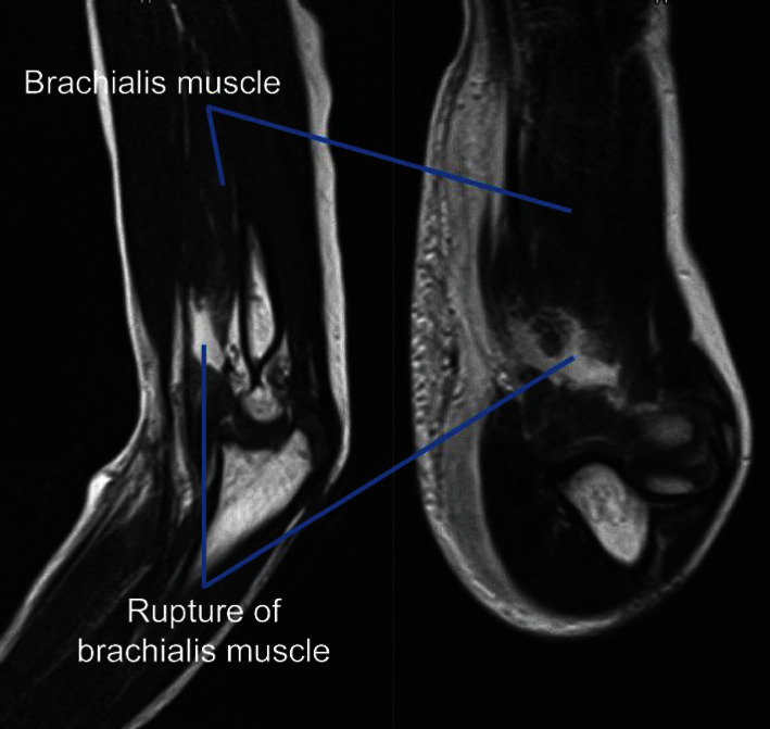
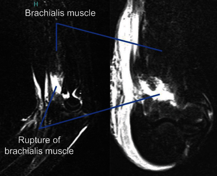
Isolated brachial muscle injuries are relatively rare injuries and reportedly occur during forced elbow extension. Though commonly conservative treatment approach is adopted, the treatment criteria remain unclear. Here, we report the case of a patient who experienced functional recovery after conservative treatment for an isolated brachial muscle injury. The patient was an 8-year-old boy whose chief complaint was left elbow pain. The injury occurred when the patient fell while playing on gymnastics bars and bruised the palmar side of his left elbow on the bar. Owing to the pain in the left elbow, the patient came to our institution. There were no clear signs of deformities or swelling in the left elbow and no obvious tenderness. X-ray and computed tomography (CT) imaging examinations revealed no signs of a fracture or dislocation, and the patient was diagnosed with left brachialis muscle rupture based on magnetic resonance imaging (MRI). Although the brachialis muscle was complete ruptured, a healing tendency was seen on body surface ultrasound examinations over time, and the patient was treated conservatively. After 3 weeks of cast immobilization, the patient underwent range of motion exercises. Two months after the injury, there were no issues with elbow joint function in daily life activities and no limitations in range of motion. Here, MRI was used to diagnose brachialis muscle rupture, and ultrasound examinations were utilized to make treatment decisions.


 求助内容:
求助内容: 应助结果提醒方式:


