Isolation efficiency of collagenase and EDTA for the culture of corneal endothelial cells.
IF 1.4 3区 医学Q4 BIOCHEMISTRY & MOLECULAR BIOLOGY
Molecular VisionPub Date : 2022-10-02eCollection Date: 2022-01-01
Kim Santerre, Stéphanie Proulx
{"title":"Isolation efficiency of collagenase and EDTA for the culture of corneal endothelial cells.","authors":"Kim Santerre, Stéphanie Proulx","doi":"","DOIUrl":null,"url":null,"abstract":"<p><strong>Purpose: </strong>Tissue engineering of the corneal endothelium, as well as cell therapy, has been proposed as an alternative approach for the treatment of corneal endotheliopathies. These approaches require in vitro amplification of functional corneal endothelial cells (CECs). The goal of this study was to compare two common isolation methods, collagenase A and EDTA (EDTA), and determine whether they influence cell viability, morphology, and barrier function.</p><p><strong>Methods: </strong>Human eye bank research-grade corneas were used to isolate and cultivate CECs. All donors were more than 40 years old. Two Descemet membranes from the same donor were used separately to compare the collagenase A and EDTA cell isolation methods. The number of isolated cells, cell viability, morphology, and barrier functionality were compared.</p><p><strong>Results: </strong>A higher isolation efficiency of viable CECs and a higher circularity index (endothelial morphology) were obtained using collagenase A. Passage 3 cells presented similar barrier functionalities regardless of the isolation method.</p><p><strong>Conclusions: </strong>This study showed that isolation of CECs using collagenase A yields higher isolation efficiency than EDTA, delaying the loss of endothelial morphology for early passage cells.</p>","PeriodicalId":18866,"journal":{"name":"Molecular Vision","volume":" ","pages":"331-339"},"PeriodicalIF":1.4000,"publicationDate":"2022-10-02","publicationTypes":"Journal Article","fieldsOfStudy":null,"isOpenAccess":false,"openAccessPdf":"https://ftp.ncbi.nlm.nih.gov/pub/pmc/oa_pdf/c6/8c/mv-v28-331.PMC9603909.pdf","citationCount":"0","resultStr":null,"platform":"Semanticscholar","paperid":null,"PeriodicalName":"Molecular Vision","FirstCategoryId":"3","ListUrlMain":"","RegionNum":3,"RegionCategory":"医学","ArticlePicture":[],"TitleCN":null,"AbstractTextCN":null,"PMCID":null,"EPubDate":"2022/1/1 0:00:00","PubModel":"eCollection","JCR":"Q4","JCRName":"BIOCHEMISTRY & MOLECULAR BIOLOGY","Score":null,"Total":0}
引用次数: 0
Abstract
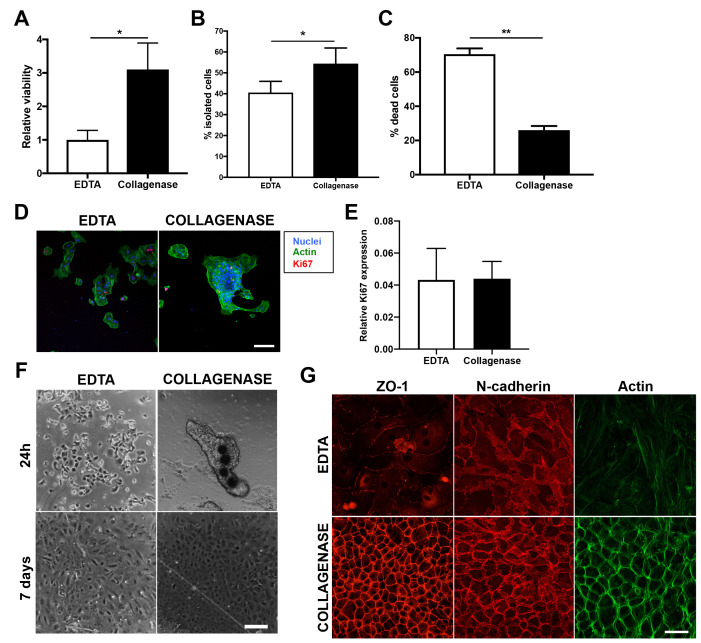
Purpose: Tissue engineering of the corneal endothelium, as well as cell therapy, has been proposed as an alternative approach for the treatment of corneal endotheliopathies. These approaches require in vitro amplification of functional corneal endothelial cells (CECs). The goal of this study was to compare two common isolation methods, collagenase A and EDTA (EDTA), and determine whether they influence cell viability, morphology, and barrier function.
Methods: Human eye bank research-grade corneas were used to isolate and cultivate CECs. All donors were more than 40 years old. Two Descemet membranes from the same donor were used separately to compare the collagenase A and EDTA cell isolation methods. The number of isolated cells, cell viability, morphology, and barrier functionality were compared.
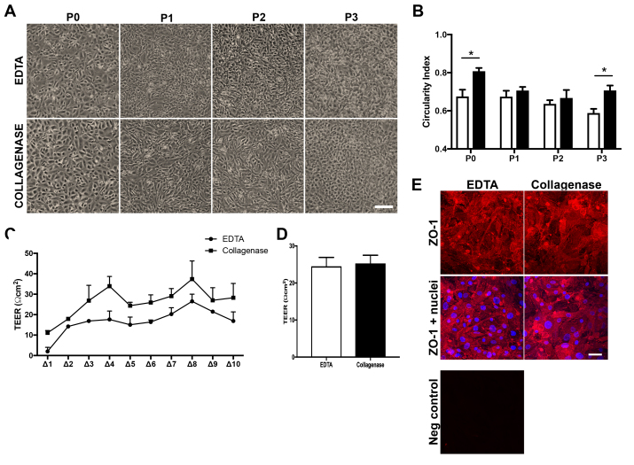
Results: A higher isolation efficiency of viable CECs and a higher circularity index (endothelial morphology) were obtained using collagenase A. Passage 3 cells presented similar barrier functionalities regardless of the isolation method.
Conclusions: This study showed that isolation of CECs using collagenase A yields higher isolation efficiency than EDTA, delaying the loss of endothelial morphology for early passage cells.
期刊介绍:
Molecular Vision is a peer-reviewed journal dedicated to the dissemination of research results in molecular biology, cell biology, and the genetics of the visual system (ocular and cortical).
Molecular Vision publishes articles presenting original research that has not previously been published and comprehensive articles reviewing the current status of a particular field or topic. Submissions to Molecular Vision are subjected to rigorous peer review. Molecular Vision does NOT publish preprints.
For authors, Molecular Vision provides a rapid means of communicating important results. Access to Molecular Vision is free and unrestricted, allowing the widest possible audience for your article. Digital publishing allows you to use color images freely (and without fees). Additionally, you may publish animations, sounds, or other supplementary information that clarifies or supports your article. Each of the authors of an article may also list an electronic mail address (which will be updated upon request) to give interested readers easy access to authors.
 求助内容:
求助内容: 应助结果提醒方式:


