Mohammad A Aziz, Benedict Seo, Haizal M Hussaini, Merilyn Hibma, Alison M Rich
{"title":"Comparing Two Methods for the Isolation of Exosomes.","authors":"Mohammad A Aziz, Benedict Seo, Haizal M Hussaini, Merilyn Hibma, Alison M Rich","doi":"10.1155/2022/8648373","DOIUrl":null,"url":null,"abstract":"<p><p>Exosomes are membrane-bound nanovesicles released by cells into their extracellular environment. They carry different types of RNA including mRNA which may be useful in the diagnosis of various diseases. Exosome isolation has been a challenge because of their small size; therefore, two exosome isolation methods were compared in this study. The Exoquick-TC PLUS™ exosome isolation kit (kit) was compared with the classic ultracentrifugation (UC) method for exosome isolation. In samples obtained using both methods, cryo-electron microscopy showed round or slightly elongated vesicles with diameters ranging from 50 to 150 nm and delimited by a bilayered membrane. Dynamic light scattering resulted in multiple peaks for kit exosomes, whereas a single peak was observed for UC exosomes. Significantly, more total RNA was present in UC exosomes in contrast to kit exosomes (<i>P</i> < 0.0001). This was reflected in subsequent mRNA analysis using qPCR, where UC exosomes had lower Ct values compared to kit exosomes. In conclusion, exosome characterization revealed the presence of exosomes in both UC and the kit samples. The kit samples presented additional peaks from DLS which might be due to impurities. Overall, due to a higher total RNA and mRNA content, UC is a better option for subsequent mRNA analysis; nevertheless, the kit can still be used if an ultracentrifuge is not available as four out of the five genes selected were detected and quantified using the kit.</p>","PeriodicalId":16575,"journal":{"name":"Journal of Nucleic Acids","volume":" ","pages":"8648373"},"PeriodicalIF":1.8000,"publicationDate":"2022-10-25","publicationTypes":"Journal Article","fieldsOfStudy":null,"isOpenAccess":false,"openAccessPdf":"https://www.ncbi.nlm.nih.gov/pmc/articles/PMC9626211/pdf/","citationCount":"1","resultStr":null,"platform":"Semanticscholar","paperid":null,"PeriodicalName":"Journal of Nucleic Acids","FirstCategoryId":"1085","ListUrlMain":"https://doi.org/10.1155/2022/8648373","RegionNum":0,"RegionCategory":null,"ArticlePicture":[],"TitleCN":null,"AbstractTextCN":null,"PMCID":null,"EPubDate":"2022/1/1 0:00:00","PubModel":"eCollection","JCR":"Q4","JCRName":"BIOCHEMISTRY & MOLECULAR BIOLOGY","Score":null,"Total":0}
引用次数: 1
Abstract
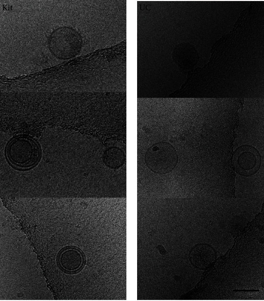
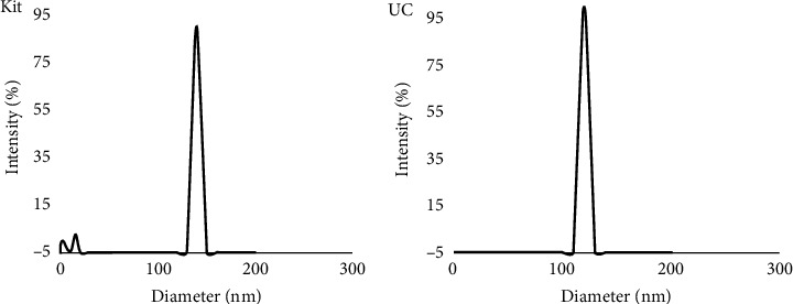
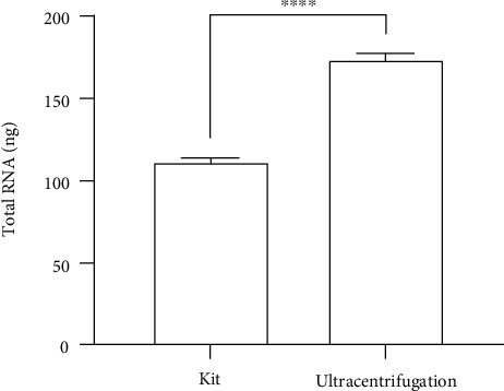
Exosomes are membrane-bound nanovesicles released by cells into their extracellular environment. They carry different types of RNA including mRNA which may be useful in the diagnosis of various diseases. Exosome isolation has been a challenge because of their small size; therefore, two exosome isolation methods were compared in this study. The Exoquick-TC PLUS™ exosome isolation kit (kit) was compared with the classic ultracentrifugation (UC) method for exosome isolation. In samples obtained using both methods, cryo-electron microscopy showed round or slightly elongated vesicles with diameters ranging from 50 to 150 nm and delimited by a bilayered membrane. Dynamic light scattering resulted in multiple peaks for kit exosomes, whereas a single peak was observed for UC exosomes. Significantly, more total RNA was present in UC exosomes in contrast to kit exosomes (P < 0.0001). This was reflected in subsequent mRNA analysis using qPCR, where UC exosomes had lower Ct values compared to kit exosomes. In conclusion, exosome characterization revealed the presence of exosomes in both UC and the kit samples. The kit samples presented additional peaks from DLS which might be due to impurities. Overall, due to a higher total RNA and mRNA content, UC is a better option for subsequent mRNA analysis; nevertheless, the kit can still be used if an ultracentrifuge is not available as four out of the five genes selected were detected and quantified using the kit.

 求助内容:
求助内容: 应助结果提醒方式:


