Xiaolong Wu, Damin Yun, Mengmeng Sang, Jianpeng Liu, Liwei Zhou, Jie Shi, Lingling Wang, Tiao Bu, Linxi Li, YingYing Huang, Dengfeng Lin, Fei Sun, C Yan Cheng
{"title":"Defects of microtubule cytoskeletal organization in NOA human testes.","authors":"Xiaolong Wu, Damin Yun, Mengmeng Sang, Jianpeng Liu, Liwei Zhou, Jie Shi, Lingling Wang, Tiao Bu, Linxi Li, YingYing Huang, Dengfeng Lin, Fei Sun, C Yan Cheng","doi":"10.1186/s12958-022-01026-w","DOIUrl":null,"url":null,"abstract":"<p><p>The importance of actin and microtubule (MT) cytoskeletons in testis function in rodents is known to some extent, but its role in the etiology of azoospermia in humans remains unexplored. Here, we examined if MT cytoskeleton was defective in NOA (non-obstructive azoospermia) testes versus normal human testes based on histopathological, immunofluorescence (IF), and scRNA-Seq transcriptome profiling. Testis biopsy samples from n = 6 normal men versus n = 3 Sertoli cell only (SCO) and n = 3 MA (meiotic arrest) of NOA patients were used for histopathological analysis. IF analysis was also used to examine MT organization across the seminiferous epithelium, investigating the likely involvement of microtubule-associated proteins (MAPs). scRNA-Seq transcriptome profiling datasets from testes of 3 SCO patients versus 3 normal men in public domain in Gene Expression Omnibus (GEO) Sample (GSM) with identifiers were analyzed to examine relevant genes that regulate MT dynamics. NOA testes of MA and SCO patients displayed notable defects in MT organization across the epithelium with extensive truncation, mis-alignments and appeared as collapsed structures near the base of the tubules. These changes are in contrast to MTs in testes of normal men. scRNA-Seq analyses revealed considerable loss of spermatogenesis capacity in SCO testes of NOA patients versus normal men. An array of genes that support MT dynamics displayed considerable changes in expression and in spatial distribution. In summary, defects in MT cytoskeleton were noted in testes of NOA (SCO) patients, possibly mediated by defective spatial expression and/or distribution of MAPs. These changes, in turn, may impede spermatogenesis in SCO testes of NOA patients.</p>","PeriodicalId":520764,"journal":{"name":"Reproductive biology and endocrinology : RB&E","volume":" ","pages":"154"},"PeriodicalIF":4.7000,"publicationDate":"2022-11-03","publicationTypes":"Journal Article","fieldsOfStudy":null,"isOpenAccess":false,"openAccessPdf":"https://www.ncbi.nlm.nih.gov/pmc/articles/PMC9632130/pdf/","citationCount":"1","resultStr":null,"platform":"Semanticscholar","paperid":null,"PeriodicalName":"Reproductive biology and endocrinology : RB&E","FirstCategoryId":"3","ListUrlMain":"https://doi.org/10.1186/s12958-022-01026-w","RegionNum":0,"RegionCategory":null,"ArticlePicture":[],"TitleCN":null,"AbstractTextCN":null,"PMCID":null,"EPubDate":"","PubModel":"","JCR":"","JCRName":"","Score":null,"Total":0}
引用次数: 1
Abstract
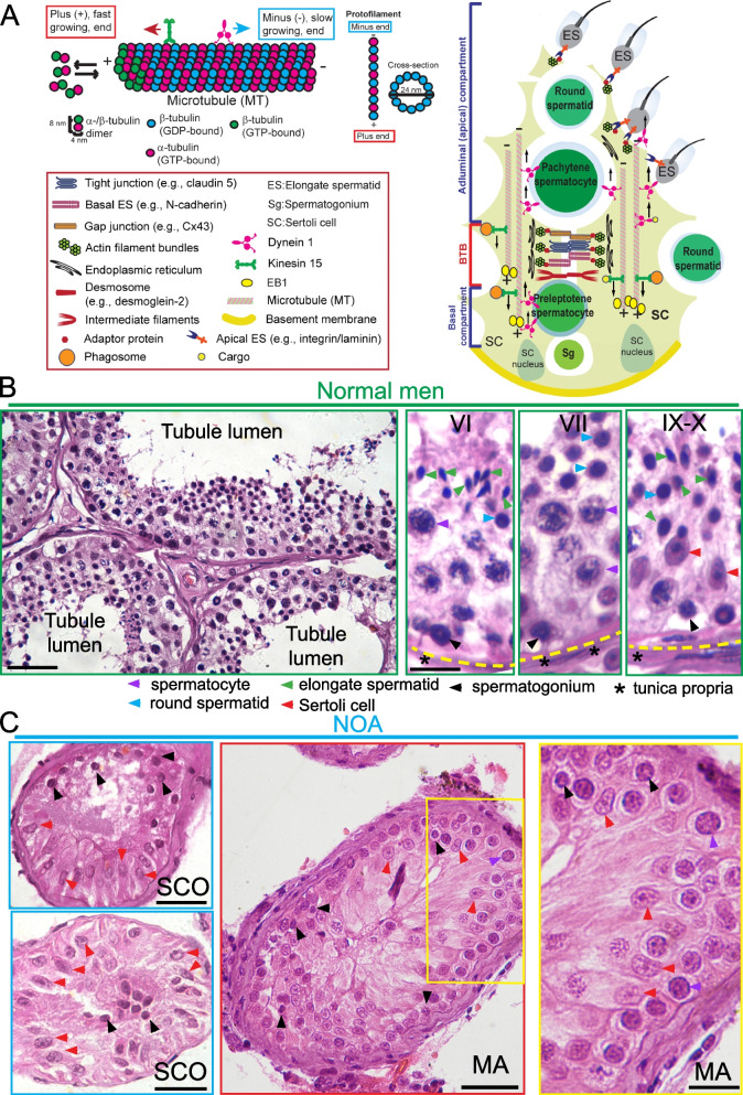
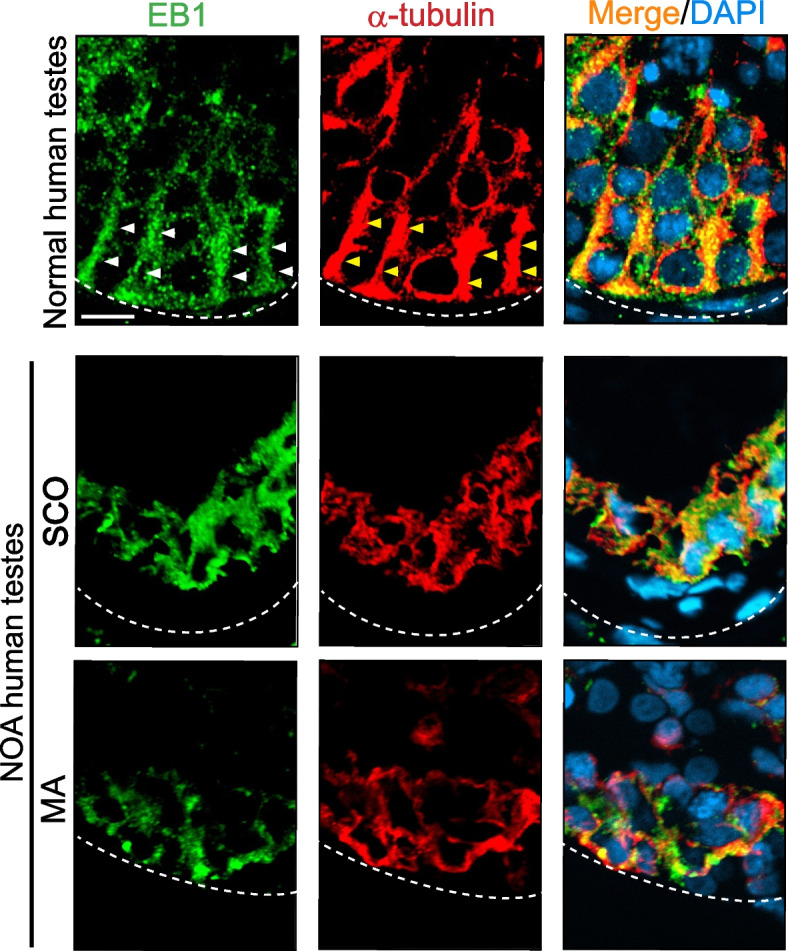
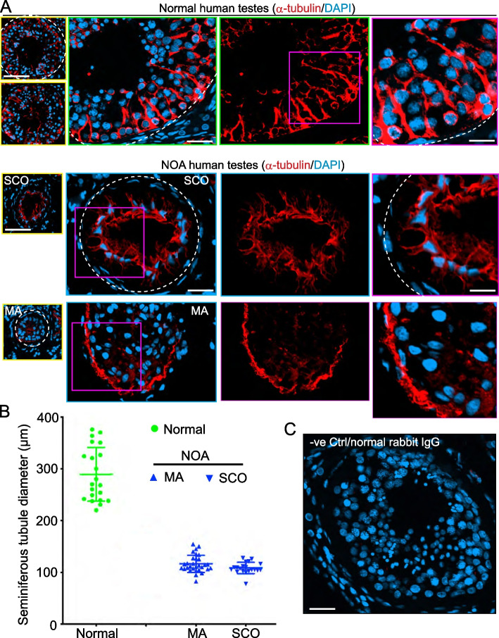
The importance of actin and microtubule (MT) cytoskeletons in testis function in rodents is known to some extent, but its role in the etiology of azoospermia in humans remains unexplored. Here, we examined if MT cytoskeleton was defective in NOA (non-obstructive azoospermia) testes versus normal human testes based on histopathological, immunofluorescence (IF), and scRNA-Seq transcriptome profiling. Testis biopsy samples from n = 6 normal men versus n = 3 Sertoli cell only (SCO) and n = 3 MA (meiotic arrest) of NOA patients were used for histopathological analysis. IF analysis was also used to examine MT organization across the seminiferous epithelium, investigating the likely involvement of microtubule-associated proteins (MAPs). scRNA-Seq transcriptome profiling datasets from testes of 3 SCO patients versus 3 normal men in public domain in Gene Expression Omnibus (GEO) Sample (GSM) with identifiers were analyzed to examine relevant genes that regulate MT dynamics. NOA testes of MA and SCO patients displayed notable defects in MT organization across the epithelium with extensive truncation, mis-alignments and appeared as collapsed structures near the base of the tubules. These changes are in contrast to MTs in testes of normal men. scRNA-Seq analyses revealed considerable loss of spermatogenesis capacity in SCO testes of NOA patients versus normal men. An array of genes that support MT dynamics displayed considerable changes in expression and in spatial distribution. In summary, defects in MT cytoskeleton were noted in testes of NOA (SCO) patients, possibly mediated by defective spatial expression and/or distribution of MAPs. These changes, in turn, may impede spermatogenesis in SCO testes of NOA patients.

 求助内容:
求助内容: 应助结果提醒方式:


