Marianne Flinck, Johan von Heideken, Ylva Aurell, Jacques Riad
{"title":"Leg length discrepancy after skeletal maturity in patients treated with elastic intramedullary nails after femoral shaft fractures in childhood.","authors":"Marianne Flinck, Johan von Heideken, Ylva Aurell, Jacques Riad","doi":"10.1177/18632521221106388","DOIUrl":null,"url":null,"abstract":"<p><strong>Purpose: </strong>The purpose was to study radiographic and perceived leg length discrepancy after skeletal maturity in patients treated for femoral shaft fractures with elastic stable intramedullary nails in childhood.</p><p><strong>Methods: </strong>Thirty-five adults underwent standing radiographs and answered a questionnaire regarding perception of leg length discrepancy. Demographic data, fracture characteristics, angulation, stability of fixation, and callus formation, at time of fracture, were assessed.</p><p><strong>Results: </strong>Mean age at fracture was 10.2 (4.9-16.7) years, and mean follow-up time was 11.1 (3.8-16.8) years. In 8 of 35 participants, the fractured limb was 11-15 mm longer than the non-fractured, and in 16, 1-10 mm longer. In eight participants, the fractured limb was 1-10 mm shorter than the non-fractured, and in three participants, 12-23 mm shorter. The younger the child, the greater the lengthening (R<sub>s</sub> = -0.49, p = 0.003). The greater the femoral angulation at time of fracture, the greater the shortening (R<sub>s</sub> = 0.42, p = 0.013). There was no significant correlation between stability of fixation or callus formation 1 month postoperatively and radiographic leg length discrepancy after skeletal maturity. Fourteen (40%) had perception of leg length discrepancy at follow-up, of whom eight had a radiographic leg length discrepancy of 10-24 mm.</p><p><strong>Conclusion: </strong>Treatment with elastic stable intramedullary nail of femoral shaft fracture in childhood may result in radiographic leg length discrepancy. Younger children were more prone to lengthening and should possibly be assessed before skeletal maturity. The degree of fracture stability or callus formation at the time of fracture did not significantly affect leg length discrepancy. Perception of leg length discrepancy was not necessarily associated with a radiographic leg length discrepancy (≥10 mm).</p><p><strong>Level of evidence: </strong>level IV, case series.</p>","PeriodicalId":138259,"journal":{"name":"Journal of Children's Orthopaedics","volume":" ","pages":"276-284"},"PeriodicalIF":0.0000,"publicationDate":"2022-08-01","publicationTypes":"Journal Article","fieldsOfStudy":null,"isOpenAccess":false,"openAccessPdf":"https://ftp.ncbi.nlm.nih.gov/pub/pmc/oa_pdf/1c/79/10.1177_18632521221106388.PMC9382705.pdf","citationCount":"2","resultStr":null,"platform":"Semanticscholar","paperid":null,"PeriodicalName":"Journal of Children's Orthopaedics","FirstCategoryId":"3","ListUrlMain":"https://doi.org/10.1177/18632521221106388","RegionNum":0,"RegionCategory":null,"ArticlePicture":[],"TitleCN":null,"AbstractTextCN":null,"PMCID":null,"EPubDate":"2022/8/2 0:00:00","PubModel":"Epub","JCR":"","JCRName":"","Score":null,"Total":0}
引用次数: 2
Abstract
Purpose: The purpose was to study radiographic and perceived leg length discrepancy after skeletal maturity in patients treated for femoral shaft fractures with elastic stable intramedullary nails in childhood.
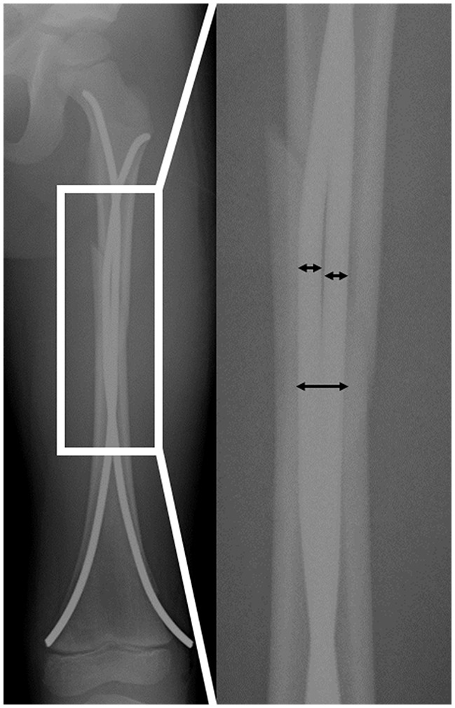
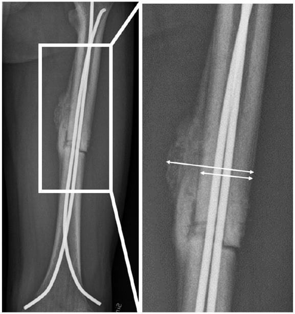
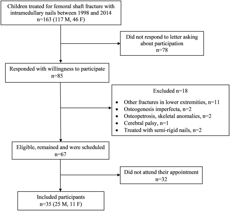
Methods: Thirty-five adults underwent standing radiographs and answered a questionnaire regarding perception of leg length discrepancy. Demographic data, fracture characteristics, angulation, stability of fixation, and callus formation, at time of fracture, were assessed.
Results: Mean age at fracture was 10.2 (4.9-16.7) years, and mean follow-up time was 11.1 (3.8-16.8) years. In 8 of 35 participants, the fractured limb was 11-15 mm longer than the non-fractured, and in 16, 1-10 mm longer. In eight participants, the fractured limb was 1-10 mm shorter than the non-fractured, and in three participants, 12-23 mm shorter. The younger the child, the greater the lengthening (Rs = -0.49, p = 0.003). The greater the femoral angulation at time of fracture, the greater the shortening (Rs = 0.42, p = 0.013). There was no significant correlation between stability of fixation or callus formation 1 month postoperatively and radiographic leg length discrepancy after skeletal maturity. Fourteen (40%) had perception of leg length discrepancy at follow-up, of whom eight had a radiographic leg length discrepancy of 10-24 mm.
Conclusion: Treatment with elastic stable intramedullary nail of femoral shaft fracture in childhood may result in radiographic leg length discrepancy. Younger children were more prone to lengthening and should possibly be assessed before skeletal maturity. The degree of fracture stability or callus formation at the time of fracture did not significantly affect leg length discrepancy. Perception of leg length discrepancy was not necessarily associated with a radiographic leg length discrepancy (≥10 mm).

 求助内容:
求助内容: 应助结果提醒方式:


