Marijn Huiskamp, Svenja Kiljan, Shanna Kulik, Maarteen E Witte, Laura E Jonkman, John Gjm Bol, Geert J Schenk, Hanneke E Hulst, Prejaas Tewarie, Menno M Schoonheim, Jeroen Jg Geurts
{"title":"Inhibitory synaptic loss drives network changes in multiple sclerosis: An ex vivo to in silico translational study.","authors":"Marijn Huiskamp, Svenja Kiljan, Shanna Kulik, Maarteen E Witte, Laura E Jonkman, John Gjm Bol, Geert J Schenk, Hanneke E Hulst, Prejaas Tewarie, Menno M Schoonheim, Jeroen Jg Geurts","doi":"10.1177/13524585221125381","DOIUrl":null,"url":null,"abstract":"<p><strong>Background: </strong>Synaptic and neuronal loss contribute to network dysfunction and disability in multiple sclerosis (MS). However, it is unknown whether excitatory or inhibitory synapses and neurons are more vulnerable and how their losses impact network functioning.</p><p><strong>Objective: </strong>To quantify excitatory and inhibitory synapses and neurons and to investigate how synaptic loss affects network functioning through computational modeling.</p><p><strong>Methods: </strong>Using immunofluorescent staining and confocal microscopy, densities of glutamatergic and GABAergic synapses and neurons were compared between post-mortem MS and non-neurological control cases. Then, a corticothalamic biophysical model was employed to study how MS-induced excitatory and inhibitory synaptic loss affect network functioning.</p><p><strong>Results: </strong>In layer VI of normal-appearing MS cortex, excitatory and inhibitory synaptic densities were significantly lower than controls (reductions up to 14.9%), but demyelinated cortex showed larger losses of inhibitory synapses (29%). In our computational model, reducing inhibitory synapses impacted the network most, leading to a disinhibitory increase in neuronal activity and connectivity.</p><p><strong>Conclusion: </strong>In MS, excitatory and inhibitory synaptic losses were observed, predominantly for inhibitory synapses in demyelinated cortex. Inhibitory synaptic loss affected network functioning most, leading to increased neuronal activity and connectivity. As network disinhibition relates to cognitive impairment, inhibitory synaptic loss seems particularly relevant in MS.</p>","PeriodicalId":520714,"journal":{"name":"Multiple sclerosis (Houndmills, Basingstoke, England)","volume":" ","pages":"2010-2019"},"PeriodicalIF":5.0000,"publicationDate":"2022-11-01","publicationTypes":"Journal Article","fieldsOfStudy":null,"isOpenAccess":false,"openAccessPdf":"https://www.ncbi.nlm.nih.gov/pmc/articles/PMC9574900/pdf/","citationCount":"5","resultStr":null,"platform":"Semanticscholar","paperid":null,"PeriodicalName":"Multiple sclerosis (Houndmills, Basingstoke, England)","FirstCategoryId":"3","ListUrlMain":"https://doi.org/10.1177/13524585221125381","RegionNum":0,"RegionCategory":null,"ArticlePicture":[],"TitleCN":null,"AbstractTextCN":null,"PMCID":null,"EPubDate":"2022/10/3 0:00:00","PubModel":"Epub","JCR":"","JCRName":"","Score":null,"Total":0}
引用次数: 5
Abstract
Background: Synaptic and neuronal loss contribute to network dysfunction and disability in multiple sclerosis (MS). However, it is unknown whether excitatory or inhibitory synapses and neurons are more vulnerable and how their losses impact network functioning.
Objective: To quantify excitatory and inhibitory synapses and neurons and to investigate how synaptic loss affects network functioning through computational modeling.
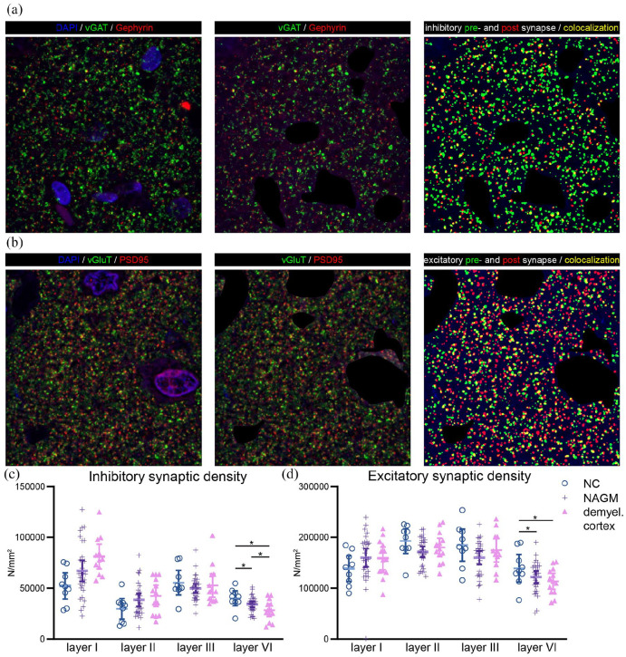
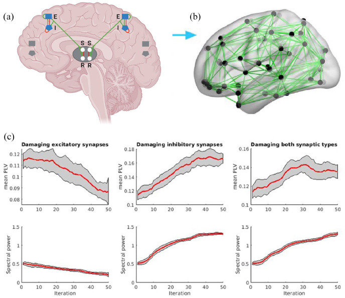
Methods: Using immunofluorescent staining and confocal microscopy, densities of glutamatergic and GABAergic synapses and neurons were compared between post-mortem MS and non-neurological control cases. Then, a corticothalamic biophysical model was employed to study how MS-induced excitatory and inhibitory synaptic loss affect network functioning.
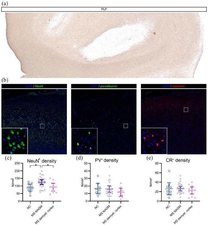
Results: In layer VI of normal-appearing MS cortex, excitatory and inhibitory synaptic densities were significantly lower than controls (reductions up to 14.9%), but demyelinated cortex showed larger losses of inhibitory synapses (29%). In our computational model, reducing inhibitory synapses impacted the network most, leading to a disinhibitory increase in neuronal activity and connectivity.
Conclusion: In MS, excitatory and inhibitory synaptic losses were observed, predominantly for inhibitory synapses in demyelinated cortex. Inhibitory synaptic loss affected network functioning most, leading to increased neuronal activity and connectivity. As network disinhibition relates to cognitive impairment, inhibitory synaptic loss seems particularly relevant in MS.

 求助内容:
求助内容: 应助结果提醒方式:


