Khaled A Obeidat, Mohammed W Afaneh, Hamzeh Mohammad Al-Domaidat, Hamzeh Ibrahim Al-Qazakzeh, Fatima J AlQaisi
{"title":"Splenic Hamartoma: A Case Report and Literature Review.","authors":"Khaled A Obeidat, Mohammed W Afaneh, Hamzeh Mohammad Al-Domaidat, Hamzeh Ibrahim Al-Qazakzeh, Fatima J AlQaisi","doi":"10.12659/AJCR.937195","DOIUrl":null,"url":null,"abstract":"<p><p>BACKGROUND Splenic hamartoma (SH) is a benign vascular lesion, usually found incidentally on abdominal images or at autopsy. Only around 200 cases have been reported since 1861, when SH was first described by Rokitansky. Although it is very rare, it is important to be familiar with it, as it may be a diagnostic challenge to distinguish SH from other mass lesions of the spleen based solely on preoperative investigations. CASE REPORT We describe a case of symptomatic, isolated, single splenic hamartoma in a 19-year-old, otherwise healthy young man who presented with upper abdominal pain, nausea, and vomiting for a few months. The examination was unremarkable. The patient has been previously evaluated with abdominal ultrasonography, which found a suspicious splenic hyperechoic lesion. Computed tomography revealed a heterogeneous 5×7 cm enhancing lesion in the spleen, concerning for splenic hamartoma. The patient underwent laparoscopic splenectomy and recovered well. The histopathology examination confirmed the diagnosis of splenic hamartoma. CONCLUSIONS Splenic hamartoma is a rare benign vascular lesion of debated etiology. Most cases are asymptomatic and are found incidentally on images, in splenectomies performed for other reasons, or at autopsy. Radiologic findings may suggest the diagnosis and new modalities have shown accuracy in distinguishing splenic hamartomas. However, resection with formal or partial splenectomy is usually still needed since the differential diagnosis is wide, from benign to aggressive lesions, and histopathology remains the criterion standard for diagnosis. Given its benign nature, we found no cases of recurrence or metastasis in the literature.</p>","PeriodicalId":205256,"journal":{"name":"The American Journal of Case Reports","volume":" ","pages":"e937195"},"PeriodicalIF":0.0000,"publicationDate":"2022-09-21","publicationTypes":"Journal Article","fieldsOfStudy":null,"isOpenAccess":false,"openAccessPdf":"https://ftp.ncbi.nlm.nih.gov/pub/pmc/oa_pdf/52/ab/amjcaserep-23-e937195.PMC9513818.pdf","citationCount":"3","resultStr":null,"platform":"Semanticscholar","paperid":null,"PeriodicalName":"The American Journal of Case Reports","FirstCategoryId":"1085","ListUrlMain":"https://doi.org/10.12659/AJCR.937195","RegionNum":0,"RegionCategory":null,"ArticlePicture":[],"TitleCN":null,"AbstractTextCN":null,"PMCID":null,"EPubDate":"","PubModel":"","JCR":"","JCRName":"","Score":null,"Total":0}
引用次数: 3
Abstract
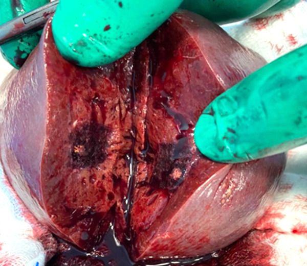
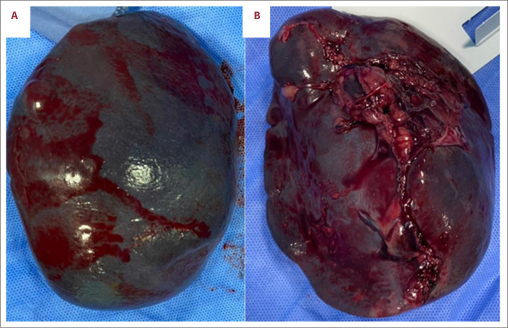
BACKGROUND Splenic hamartoma (SH) is a benign vascular lesion, usually found incidentally on abdominal images or at autopsy. Only around 200 cases have been reported since 1861, when SH was first described by Rokitansky. Although it is very rare, it is important to be familiar with it, as it may be a diagnostic challenge to distinguish SH from other mass lesions of the spleen based solely on preoperative investigations. CASE REPORT We describe a case of symptomatic, isolated, single splenic hamartoma in a 19-year-old, otherwise healthy young man who presented with upper abdominal pain, nausea, and vomiting for a few months. The examination was unremarkable. The patient has been previously evaluated with abdominal ultrasonography, which found a suspicious splenic hyperechoic lesion. Computed tomography revealed a heterogeneous 5×7 cm enhancing lesion in the spleen, concerning for splenic hamartoma. The patient underwent laparoscopic splenectomy and recovered well. The histopathology examination confirmed the diagnosis of splenic hamartoma. CONCLUSIONS Splenic hamartoma is a rare benign vascular lesion of debated etiology. Most cases are asymptomatic and are found incidentally on images, in splenectomies performed for other reasons, or at autopsy. Radiologic findings may suggest the diagnosis and new modalities have shown accuracy in distinguishing splenic hamartomas. However, resection with formal or partial splenectomy is usually still needed since the differential diagnosis is wide, from benign to aggressive lesions, and histopathology remains the criterion standard for diagnosis. Given its benign nature, we found no cases of recurrence or metastasis in the literature.


 求助内容:
求助内容: 应助结果提醒方式:


