Jia Liu, Lei Liu, Yang Su, Yi Wang, Yuchun Zhu, Xiaobin Sun, Yuanbiao Guo, Jing Shan
{"title":"IL-33 Participates in the Development of Esophageal Adenocarcinoma.","authors":"Jia Liu, Lei Liu, Yang Su, Yi Wang, Yuchun Zhu, Xiaobin Sun, Yuanbiao Guo, Jing Shan","doi":"10.3389/pore.2022.1610474","DOIUrl":null,"url":null,"abstract":"<p><p><b>Background:</b> The progression from chronic gastroesophageal reflux disease (GERD) to Barrett esophagus (BE) and esophageal adenocarcinoma (EAC) is an inflammatory-driven neoplastic change. Interleukin-33 (IL-33) has identified as a crucial factor in several inflammatory disorders and malignancies. <b>Methods:</b> The high-density tissue microarray of the human EAC was analyzed with IL-33 immunohistochemistry staining (IHC). By anastomosing the jejunum with the esophagus, the rat model of EAC with mixed gastroduodenal reflux was established. The expression of IL-33 was determined using quantitative real-time polymerase chain reaction (RT-qPCR), western blot (WB), IHC and enzyme-linked immunosorbent assay (ELISA). Esophageal adenocarcinoma cells (OE19 and OE33) and human esophageal epithelial cells (HEECs) were used. <b>Results:</b> In the cytoplasm of human EAC tissue, IL-33 expression was substantially greater than in adjacent normal tissue. In rat model, the expression of IL-33 in the EAC group was considerably greater than in the control group, and this expression increased with the upgrade of pathological stage. In <i>in vitro</i> experiment, the mRNA and protein levels of IL-33 were considerably greater in OE19 and OE33 than in HEECs. The stimulation of IL-33 enhanced the proliferation, migration, invasion, and epithelial-mesenchymal transition (EMT) of OE19 and OE33, but soluble ST2 (sST2) inhibited these effects. IL-33 stimulated the release of IL-6 by OE19 and OE33 cells. <b>Conclusion:</b> This study demonstrated the overexpression of IL-33 in the transition from GERD to EAC and that IL-33 promoted carcinogenesis in EAC cells through ST2. IL-33 might be a possible preventive target for EAC.</p>","PeriodicalId":411887,"journal":{"name":"Pathology oncology research : POR","volume":" ","pages":"1610474"},"PeriodicalIF":0.0000,"publicationDate":"2022-08-30","publicationTypes":"Journal Article","fieldsOfStudy":null,"isOpenAccess":false,"openAccessPdf":"https://www.ncbi.nlm.nih.gov/pmc/articles/PMC9469785/pdf/","citationCount":"2","resultStr":null,"platform":"Semanticscholar","paperid":null,"PeriodicalName":"Pathology oncology research : POR","FirstCategoryId":"3","ListUrlMain":"https://doi.org/10.3389/pore.2022.1610474","RegionNum":0,"RegionCategory":null,"ArticlePicture":[],"TitleCN":null,"AbstractTextCN":null,"PMCID":null,"EPubDate":"2022/1/1 0:00:00","PubModel":"eCollection","JCR":"","JCRName":"","Score":null,"Total":0}
引用次数: 2
Abstract
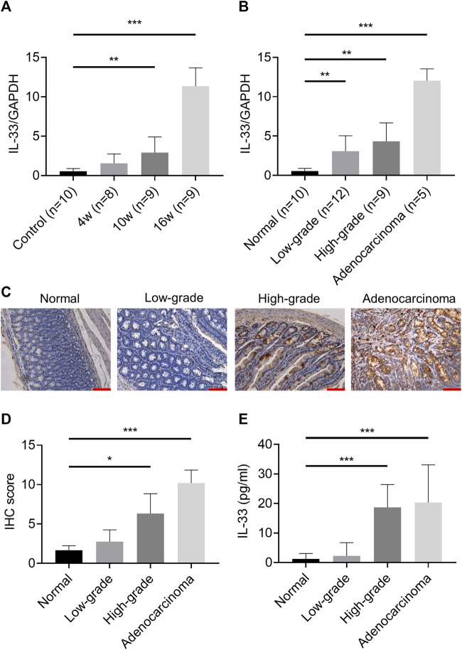
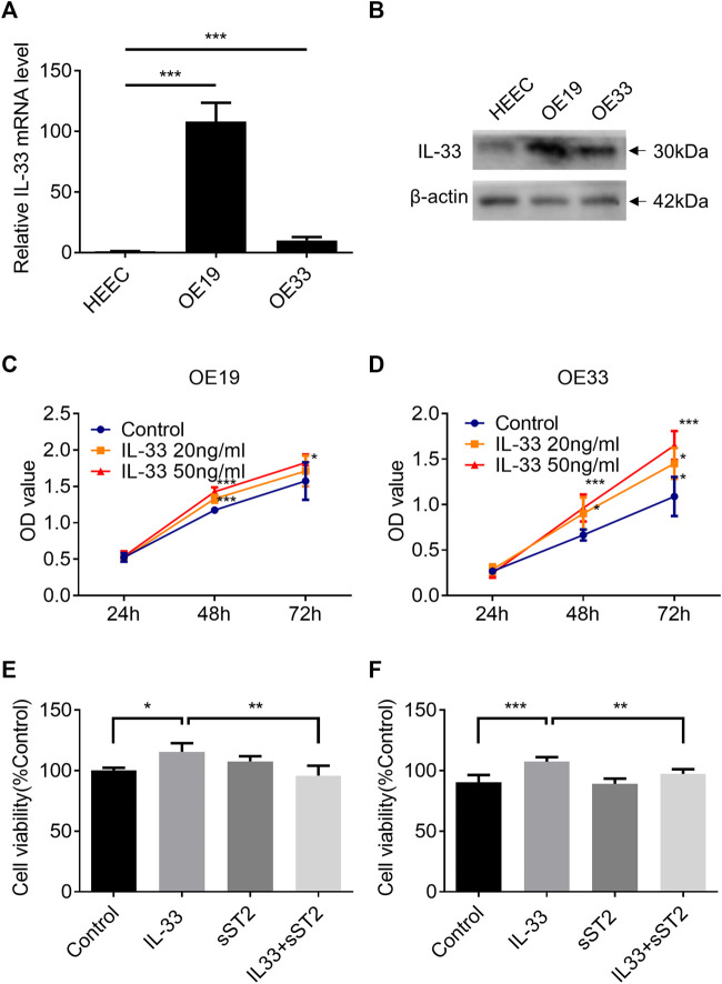
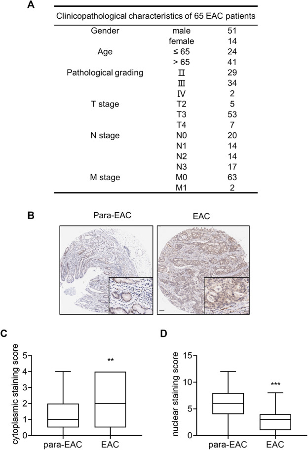
Background: The progression from chronic gastroesophageal reflux disease (GERD) to Barrett esophagus (BE) and esophageal adenocarcinoma (EAC) is an inflammatory-driven neoplastic change. Interleukin-33 (IL-33) has identified as a crucial factor in several inflammatory disorders and malignancies. Methods: The high-density tissue microarray of the human EAC was analyzed with IL-33 immunohistochemistry staining (IHC). By anastomosing the jejunum with the esophagus, the rat model of EAC with mixed gastroduodenal reflux was established. The expression of IL-33 was determined using quantitative real-time polymerase chain reaction (RT-qPCR), western blot (WB), IHC and enzyme-linked immunosorbent assay (ELISA). Esophageal adenocarcinoma cells (OE19 and OE33) and human esophageal epithelial cells (HEECs) were used. Results: In the cytoplasm of human EAC tissue, IL-33 expression was substantially greater than in adjacent normal tissue. In rat model, the expression of IL-33 in the EAC group was considerably greater than in the control group, and this expression increased with the upgrade of pathological stage. In in vitro experiment, the mRNA and protein levels of IL-33 were considerably greater in OE19 and OE33 than in HEECs. The stimulation of IL-33 enhanced the proliferation, migration, invasion, and epithelial-mesenchymal transition (EMT) of OE19 and OE33, but soluble ST2 (sST2) inhibited these effects. IL-33 stimulated the release of IL-6 by OE19 and OE33 cells. Conclusion: This study demonstrated the overexpression of IL-33 in the transition from GERD to EAC and that IL-33 promoted carcinogenesis in EAC cells through ST2. IL-33 might be a possible preventive target for EAC.

 求助内容:
求助内容: 应助结果提醒方式:


