Marek Porowski, Henryk Skarżyński, Piotr Henryk Skarzynski
{"title":"Stapedotomy in Congenital Stapes Ankylosis with Mobile Footplate: A Case Report.","authors":"Marek Porowski, Henryk Skarżyński, Piotr Henryk Skarzynski","doi":"10.12659/AJCR.936466","DOIUrl":null,"url":null,"abstract":"<p><p>BACKGROUND This paper presents a case of a 15-year-old child with a rare congenital anomaly of the middle ear in which the stapes was fused to the medial wall of the tympanic cavity. CASE REPORT This defect coexisted with partial malleus fixation in the attic and caused conductive hearing loss at an average level of 35-40 dB. Two exploratory tympanotomies were performed, where excess bone between the stapes and promontory was removed and the head of the malleus was released in the attic. The good effect of these procedures was unstable, mainly due to re-attachment of the stapes to the medial wall of the tympanic cavity. At the next surgery it was decided to perform stapedotomy, despite the mobile stapes footplate. The operation was performed with a small-fenestra stapedotomy technique. Perforation of the footplate was done using a microdrill with a balanced speed. A KURZ prosthesis with a diameter of 0.5 mm was used. The postoperative period passed without any complications. Three and 6 months after the surgery, control pure tone audiometry was performed and showed significant improvement in hearing thresholds. During the follow-up period of more than 6 months, hearing improvement remained stable. CONCLUSIONS We concluded that it was safe to perform stapedotomy in the presence of a mobile stapes footplate when congenital anomaly of the stapes superstructure caused its severe fixation in the middle ear. In our opinion, in a child with congenital ear anomaly, consideration should be given to the multifocal origin of the hearing loss.</p>","PeriodicalId":205256,"journal":{"name":"The American Journal of Case Reports","volume":" ","pages":"e936466"},"PeriodicalIF":0.0000,"publicationDate":"2022-09-15","publicationTypes":"Journal Article","fieldsOfStudy":null,"isOpenAccess":false,"openAccessPdf":"https://ftp.ncbi.nlm.nih.gov/pub/pmc/oa_pdf/b8/cc/amjcaserep-23-e936466.PMC9483239.pdf","citationCount":"0","resultStr":null,"platform":"Semanticscholar","paperid":null,"PeriodicalName":"The American Journal of Case Reports","FirstCategoryId":"1085","ListUrlMain":"https://doi.org/10.12659/AJCR.936466","RegionNum":0,"RegionCategory":null,"ArticlePicture":[],"TitleCN":null,"AbstractTextCN":null,"PMCID":null,"EPubDate":"","PubModel":"","JCR":"","JCRName":"","Score":null,"Total":0}
引用次数: 0
Abstract
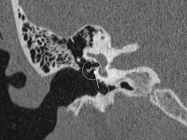
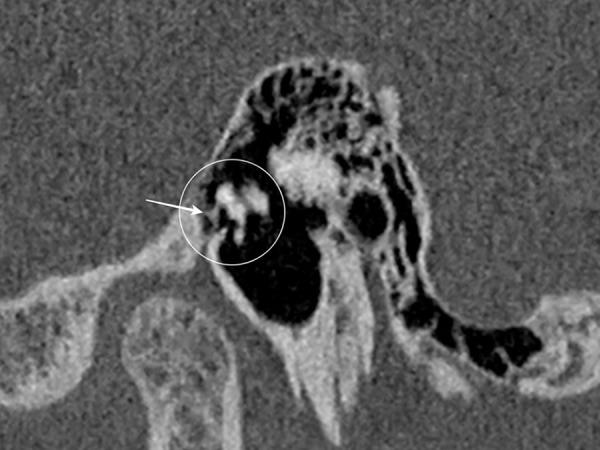
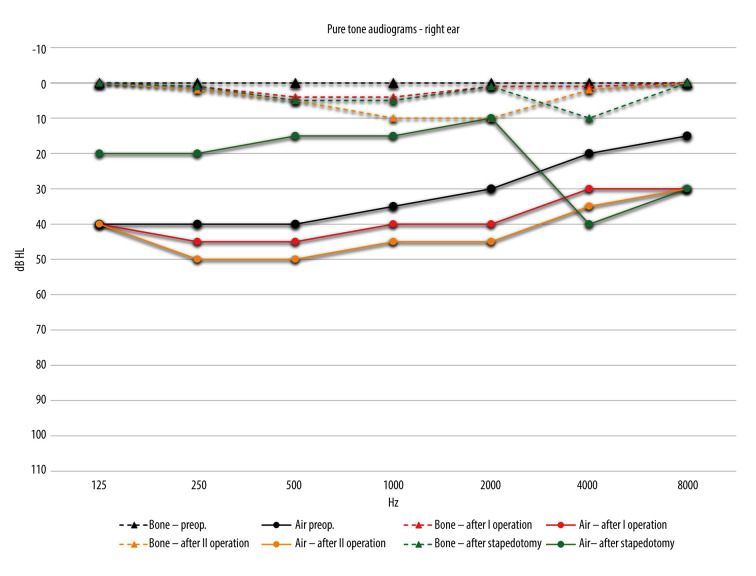
BACKGROUND This paper presents a case of a 15-year-old child with a rare congenital anomaly of the middle ear in which the stapes was fused to the medial wall of the tympanic cavity. CASE REPORT This defect coexisted with partial malleus fixation in the attic and caused conductive hearing loss at an average level of 35-40 dB. Two exploratory tympanotomies were performed, where excess bone between the stapes and promontory was removed and the head of the malleus was released in the attic. The good effect of these procedures was unstable, mainly due to re-attachment of the stapes to the medial wall of the tympanic cavity. At the next surgery it was decided to perform stapedotomy, despite the mobile stapes footplate. The operation was performed with a small-fenestra stapedotomy technique. Perforation of the footplate was done using a microdrill with a balanced speed. A KURZ prosthesis with a diameter of 0.5 mm was used. The postoperative period passed without any complications. Three and 6 months after the surgery, control pure tone audiometry was performed and showed significant improvement in hearing thresholds. During the follow-up period of more than 6 months, hearing improvement remained stable. CONCLUSIONS We concluded that it was safe to perform stapedotomy in the presence of a mobile stapes footplate when congenital anomaly of the stapes superstructure caused its severe fixation in the middle ear. In our opinion, in a child with congenital ear anomaly, consideration should be given to the multifocal origin of the hearing loss.

 求助内容:
求助内容: 应助结果提醒方式:


