Quantitative Analysis in Cervical Spinal Cord Injury Patients Using Diffusion Tensor Imaging and Tractography.
引用次数: 0
Abstract
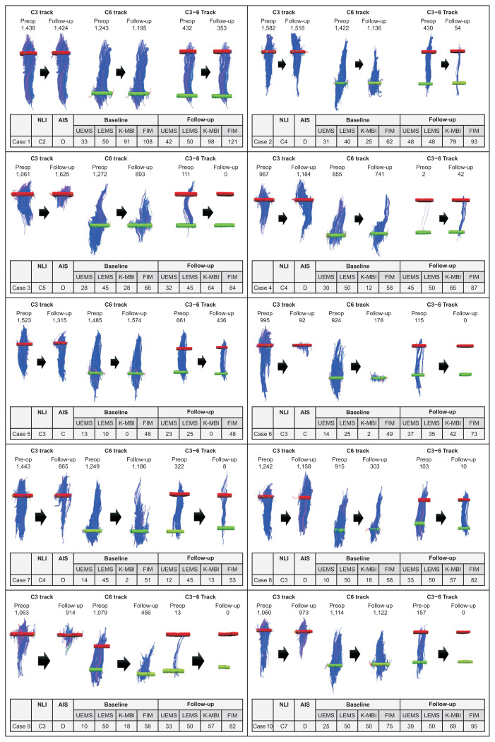
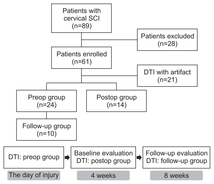
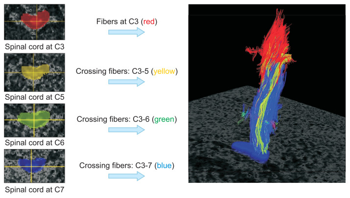
Objective To investigate the clinical usefulness of diffusion tensor imaging (DTI) and tractography in the prediction of outcomes after traumatic cervical spinal cord injury (SCI) and to assess whether the predictability is different between DTI and tractography administered before and after surgery. Methods Sixty-one subjects with traumatic cervical SCI were randomly assigned to preop or postop groups and received DTI accordingly. Among the patients who had DTI before surgery, we assigned 10 patients who had received repeated DTI examinations at 8 weeks after injury to the follow-up group. Fractional anisotropy (FA) and apparent diffusion coefficient (ADC) values were obtained from DTI, and imaginary fiber and crossing fiber numbers were calculated from the tractography. Neurological status and functional status were assessed at 4 and 8 weeks after SCI. Results The neurologic and functional statuses of both groups improved after 4 weeks. Out of the initial 61 patients who were enrolled in the study, the failure rate of DTI image analysis was significantly higher in the postop group (n=17, 41.5%) than in the preop group (n=6, 20%). The FA values and fiber numbers in the preop group tended to be higher than those in the postop group, whereas ADC values were lower in the preop group. When comparing the tractographic findings in the follow-up group, imaginary fiber numbers at the C6 and C7 levels and crossing fiber numbers from the C3 to C6 levels were significantly decreased after surgery. Several DTI and tractographic parameters (especially the ADC value at the C4 level and imaginary fiber numbers at the C6 level) showed significant correlations with neurologic and functional statuses in both the preop and postop groups. These findings were most prominent when DTI and physical examination were simultaneously performed. Conclusion Preoperative DTI and tractography demonstrated better FA and ADC values with lower interpretation failure rates than those obtained after surgery, whereas postoperative data significantly reflected the patient’s clinical state at the time of evaluation. Therefore, DTI and tractography could be useful in predicting clinical outcomes after traumatic cervical SCI and should be interpreted separately before and after spine surgery.
应用弥散张量成像和脊髓束造影定量分析颈脊髓损伤患者。
目的:探讨弥散张量成像(diffusion tensor imaging, DTI)和神经束造影(tractography)在预测外伤性颈脊髓损伤(SCI)预后中的临床应用价值,并评估术前和术后DTI和神经束造影在预测预后方面是否存在差异。方法:将61例外伤性颈椎损伤患者随机分为手术前组和手术后组,分别进行DTI治疗。在术前有DTI的患者中,我们将10例在损伤后8周反复接受DTI检查的患者分配到随访组。通过DTI得到分数各向异性(FA)和表观扩散系数(ADC),通过束束成像计算虚纤维数和交叉纤维数。脊髓损伤后4周和8周评估神经状态和功能状态。结果:治疗4周后,两组患者的神经功能状况均有改善。在最初纳入研究的61例患者中,术后组的DTI图像分析失败率(n= 17,41.5%)明显高于术前组(n= 6,20%)。术前组FA值和纤维数高于术后组,ADC值低于术前组。与随访组的纤维束造影结果相比,术后C6和C7节段的虚纤维数以及C3到C6节段的交叉纤维数明显减少。术前和术后的DTI和牵道图参数(特别是C4水平的ADC值和C6水平的虚纤维数)与神经和功能状态均有显著相关性。当DTI和体格检查同时进行时,这些发现最为突出。结论:术前DTI和肛管造影比术后FA和ADC值更好,解释失败率更低,而术后数据显著反映了评估时患者的临床状态。因此,DTI和脊髓束造影可用于预测外伤性颈椎脊髓损伤后的临床结果,应在脊柱手术前后分别进行分析。
本文章由计算机程序翻译,如有差异,请以英文原文为准。
求助全文
约1分钟内获得全文
求助全文

 求助内容:
求助内容: 应助结果提醒方式:


