Conservation and divergence between cytoplasmic and muscle-specific actin capping proteins: insights from the crystal structure of cytoplasmic Cap32/34 from Dictyostelium discoideum
Christian Eckert, Agnieszka Goretzki, Maria Faberova, Martin Kollmar
{"title":"Conservation and divergence between cytoplasmic and muscle-specific actin capping proteins: insights from the crystal structure of cytoplasmic Cap32/34 from Dictyostelium discoideum","authors":"Christian Eckert, Agnieszka Goretzki, Maria Faberova, Martin Kollmar","doi":"10.1186/1472-6807-12-12","DOIUrl":null,"url":null,"abstract":"<p>Capping protein (CP), also known as CapZ in muscle cells and Cap32/34 in <i>Dictyostelium discoideum</i>, plays a major role in regulating actin filament dynamics. CP is a ubiquitously expressed heterodimer comprising an α- and β-subunit. It tightly binds to the fast growing end of actin filaments, thereby functioning as a “cap” by blocking the addition and loss of actin subunits. Vertebrates contain two somatic variants of CP, one being primarily found at the cell periphery of non-muscle tissues while the other is mainly localized at the Z-discs of skeletal muscles.</p><p>To elucidate structural and functional differences between cytoplasmic and sarcomercic CP variants, we have solved the atomic structure of Cap32/34 (32?=?β- and 34?=?α-subunit) from the cellular slime mold <i>Dictyostelium</i> at 2.2?? resolution and compared it to that of chicken muscle CapZ. The two homologs display a similar overall arrangement including the attached α-subunit C-terminus (α-tentacle) and the flexible β-tentacle. Nevertheless, the structures exhibit marked differences suggesting considerable structural flexibility within the α-subunit. In the α-subunit we observed a bending motion of the β-sheet region located opposite to the position of the C-terminal β-tentacle towards the antiparallel helices that interconnect the heterodimer. Recently, a two domain twisting attributed mainly to the β-subunit has been reported. At the hinge of these two domains Cap32/34 contains an elongated and highly flexible loop, which has been reported to be important for the interaction of cytoplasmic CP with actin and might contribute to the more dynamic actin-binding of cytoplasmic compared to sarcomeric CP (CapZ).</p><p>The structure of Cap32/34 from <i>Dictyostelium discoideum</i> allowed a detailed analysis and comparison between the cytoplasmic and sarcomeric variants of CP. Significant structural flexibility could particularly be found within the α-subunit, a loop region in the β-subunit, and the surface of the α-globule where the amino acid differences between the cytoplasmic and sarcomeric mammalian CP are located. Hence, the crystal structure of Cap32/34 raises the possibility of different binding behaviours of the CP variants toward the barbed end of actin filaments, a feature, which might have arisen from adaptation to different environments.</p>","PeriodicalId":51240,"journal":{"name":"BMC Structural Biology","volume":"12 1","pages":""},"PeriodicalIF":0.0000,"publicationDate":"2012-06-01","publicationTypes":"Journal Article","fieldsOfStudy":null,"isOpenAccess":false,"openAccessPdf":"https://sci-hub-pdf.com/10.1186/1472-6807-12-12","citationCount":"4","resultStr":null,"platform":"Semanticscholar","paperid":null,"PeriodicalName":"BMC Structural Biology","FirstCategoryId":"1085","ListUrlMain":"https://link.springer.com/article/10.1186/1472-6807-12-12","RegionNum":0,"RegionCategory":null,"ArticlePicture":[],"TitleCN":null,"AbstractTextCN":null,"PMCID":null,"EPubDate":"","PubModel":"","JCR":"Q3","JCRName":"Biochemistry, Genetics and Molecular Biology","Score":null,"Total":0}
引用次数: 4
Abstract
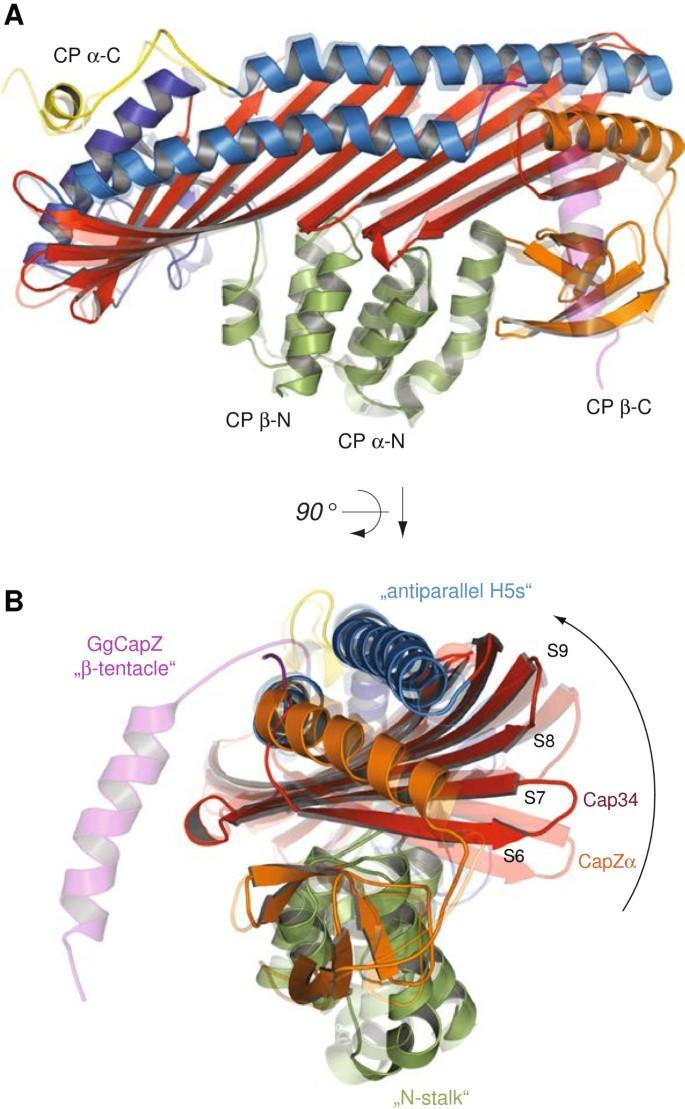
Capping protein (CP), also known as CapZ in muscle cells and Cap32/34 in Dictyostelium discoideum, plays a major role in regulating actin filament dynamics. CP is a ubiquitously expressed heterodimer comprising an α- and β-subunit. It tightly binds to the fast growing end of actin filaments, thereby functioning as a “cap” by blocking the addition and loss of actin subunits. Vertebrates contain two somatic variants of CP, one being primarily found at the cell periphery of non-muscle tissues while the other is mainly localized at the Z-discs of skeletal muscles.
To elucidate structural and functional differences between cytoplasmic and sarcomercic CP variants, we have solved the atomic structure of Cap32/34 (32?=?β- and 34?=?α-subunit) from the cellular slime mold Dictyostelium at 2.2?? resolution and compared it to that of chicken muscle CapZ. The two homologs display a similar overall arrangement including the attached α-subunit C-terminus (α-tentacle) and the flexible β-tentacle. Nevertheless, the structures exhibit marked differences suggesting considerable structural flexibility within the α-subunit. In the α-subunit we observed a bending motion of the β-sheet region located opposite to the position of the C-terminal β-tentacle towards the antiparallel helices that interconnect the heterodimer. Recently, a two domain twisting attributed mainly to the β-subunit has been reported. At the hinge of these two domains Cap32/34 contains an elongated and highly flexible loop, which has been reported to be important for the interaction of cytoplasmic CP with actin and might contribute to the more dynamic actin-binding of cytoplasmic compared to sarcomeric CP (CapZ).
The structure of Cap32/34 from Dictyostelium discoideum allowed a detailed analysis and comparison between the cytoplasmic and sarcomeric variants of CP. Significant structural flexibility could particularly be found within the α-subunit, a loop region in the β-subunit, and the surface of the α-globule where the amino acid differences between the cytoplasmic and sarcomeric mammalian CP are located. Hence, the crystal structure of Cap32/34 raises the possibility of different binding behaviours of the CP variants toward the barbed end of actin filaments, a feature, which might have arisen from adaptation to different environments.
期刊介绍:
BMC Structural Biology is an open access, peer-reviewed journal that considers articles on investigations into the structure of biological macromolecules, including solving structures, structural and functional analyses, and computational modeling.

 求助内容:
求助内容: 应助结果提醒方式:


