{"title":"Needle aspirate PTH in diagnosis of primary hyperparathyroidism due to intrathyroidal parathyroid cyst.","authors":"Deep Dutta, Chitra Selvan, Manoj Kumar, Saumik Datta, Ram Narayan Das, Sujoy Ghosh, Satinath Mukhopadhyay, Subhankar Chowdhury","doi":"10.1530/EDM-13-0019","DOIUrl":null,"url":null,"abstract":"<p><strong>Unlabelled: </strong>Parathyroid cysts are rare (0.8-3.41% of all parathyroid lesions) and usually arise secondary to cystic degeneration of parathyroid adenomas. Intrathyroidal parathyroid cysts are extremely rare with only three cases reported till date. We present a 24-year-old female with clinical and biochemical features of primary hyperparathyroidism (PHPT; Ca(2) (+): 12.1 mg/dl; intact parathyroid hormone (iPTH): 1283 pg/ml) and poor radiotracer uptake with minimal residual uptake in the left thyroid lobe at 2 and 4 h on Tc(99m) sestamibi imaging. Neck ultrasonography (USG) revealed 0.6×1 cm parathyroid posterior left lobe of thyroid along with 22×18 mm simple thyroid cyst. USG-guided fine-needle aspiration (FNA) and needle tip iPTH estimation (FNA-iPTH) from parathyroid lesion was inconclusive (114 pg/ml), necessitating FNA of thyroid cyst, which revealed high iPTH (3480 pg/ml) from the aspirate. The patient underwent a left hemithyroidectomy. A >50% drop in serum iPTH 20 min after left hemithyroidectomy (29.4 pg/ml) along with histopathology suggestive of intrathyroidal cystic parathyroid adenoma (cystic lesion lined by chief cell variant parathyroid cells without any nuclear atypia, capsular or vascular invasion surrounded by normal thyroid follicles) confirmed that the parathyroid cyst was responsible for PHPT. This report highlights the importance of FNA-iPTH in localizing and differentiating a functional parathyroid lesion from nonfunctional tissue in PHPT.</p><p><strong>Learning points: </strong>Fine-needle aspiration from suspected parathyroid lesion and needle tip iPTH (FNA-iPTH) estimation from the saline washing has an important role in localizing primary hyperparathyroidism (PHPT).FNA-iPTH estimation may help in differentiating functional from nonfunctional parathyroid lesion responsible for PHPT.iPTH estimation from aspirate of an intrathyroid cyst is helpful in differentiating intrathyroidal parathyroid cyst from thyroid cyst.</p>","PeriodicalId":520608,"journal":{"name":"Endocrinology, diabetes & metabolism case reports","volume":" ","pages":"130019"},"PeriodicalIF":0.7000,"publicationDate":"2013-01-01","publicationTypes":"Journal Article","fieldsOfStudy":null,"isOpenAccess":false,"openAccessPdf":"https://www.ncbi.nlm.nih.gov/pmc/articles/PMC3922279/pdf/","citationCount":"18","resultStr":null,"platform":"Semanticscholar","paperid":null,"PeriodicalName":"Endocrinology, diabetes & metabolism case reports","FirstCategoryId":"1085","ListUrlMain":"https://doi.org/10.1530/EDM-13-0019","RegionNum":0,"RegionCategory":null,"ArticlePicture":[],"TitleCN":null,"AbstractTextCN":null,"PMCID":null,"EPubDate":"2013/7/1 0:00:00","PubModel":"Epub","JCR":"","JCRName":"","Score":null,"Total":0}
引用次数: 18
Abstract
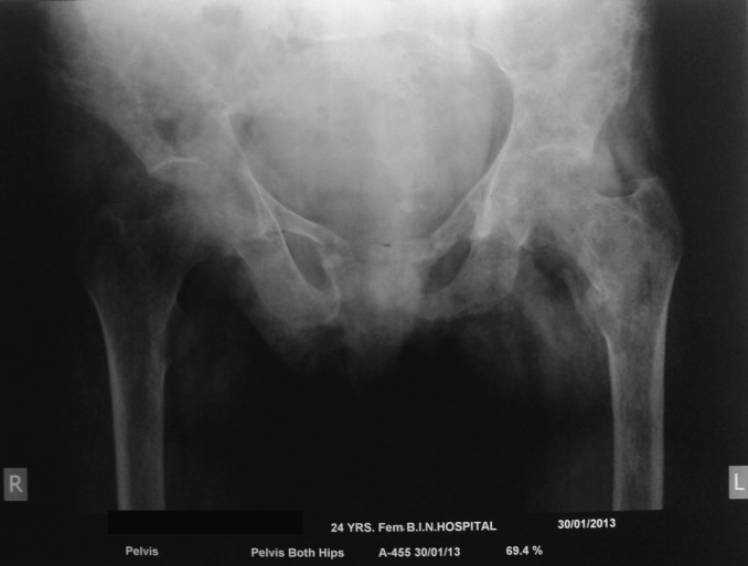
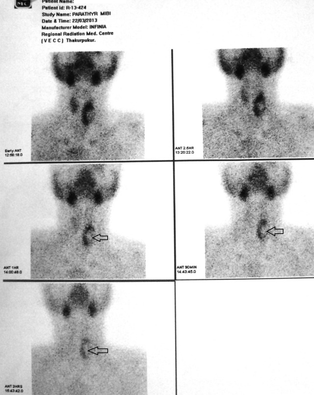
Unlabelled: Parathyroid cysts are rare (0.8-3.41% of all parathyroid lesions) and usually arise secondary to cystic degeneration of parathyroid adenomas. Intrathyroidal parathyroid cysts are extremely rare with only three cases reported till date. We present a 24-year-old female with clinical and biochemical features of primary hyperparathyroidism (PHPT; Ca(2) (+): 12.1 mg/dl; intact parathyroid hormone (iPTH): 1283 pg/ml) and poor radiotracer uptake with minimal residual uptake in the left thyroid lobe at 2 and 4 h on Tc(99m) sestamibi imaging. Neck ultrasonography (USG) revealed 0.6×1 cm parathyroid posterior left lobe of thyroid along with 22×18 mm simple thyroid cyst. USG-guided fine-needle aspiration (FNA) and needle tip iPTH estimation (FNA-iPTH) from parathyroid lesion was inconclusive (114 pg/ml), necessitating FNA of thyroid cyst, which revealed high iPTH (3480 pg/ml) from the aspirate. The patient underwent a left hemithyroidectomy. A >50% drop in serum iPTH 20 min after left hemithyroidectomy (29.4 pg/ml) along with histopathology suggestive of intrathyroidal cystic parathyroid adenoma (cystic lesion lined by chief cell variant parathyroid cells without any nuclear atypia, capsular or vascular invasion surrounded by normal thyroid follicles) confirmed that the parathyroid cyst was responsible for PHPT. This report highlights the importance of FNA-iPTH in localizing and differentiating a functional parathyroid lesion from nonfunctional tissue in PHPT.
Learning points: Fine-needle aspiration from suspected parathyroid lesion and needle tip iPTH (FNA-iPTH) estimation from the saline washing has an important role in localizing primary hyperparathyroidism (PHPT).FNA-iPTH estimation may help in differentiating functional from nonfunctional parathyroid lesion responsible for PHPT.iPTH estimation from aspirate of an intrathyroid cyst is helpful in differentiating intrathyroidal parathyroid cyst from thyroid cyst.


 求助内容:
求助内容: 应助结果提醒方式:


