Sonja E Steigen, Niels Ramsing Holm, Noreen Butt, Michael Maeng, Fumiyuki Otsuka, Renu Virmani, Elena Ladich, Terje K Steigen
{"title":"Clinical use of optical coherence tomography to identify angiographic silent stent thrombosis.","authors":"Sonja E Steigen, Niels Ramsing Holm, Noreen Butt, Michael Maeng, Fumiyuki Otsuka, Renu Virmani, Elena Ladich, Terje K Steigen","doi":"10.3109/14017431.2014.900185","DOIUrl":null,"url":null,"abstract":"<p><strong>Objectives: </strong>Patients previously treated with coronary stents may suffer an acute coronary syndrome (ACS) without any evidence of thrombus formation on coronary angiography (CAG). This may be due to partial, nonocclusive stent thrombosis with microembolization. In this paper, we illustrate possible mechanisms both with optical coherence tomography (OCT) and histology.</p><p><strong>Design: </strong>We present two cases with ACS from very late stent thrombosis who have been previously treated with first-generation drug-eluting stents (DES).</p><p><strong>Results: </strong>The first patient had ACS 15 months after DES implantation. The angiogram (CAG) was near normal with slight peri-stent contrast staining. OCT revealed abnormalities including thrombus not visible on CAG. These are findings that may explain the ACS. The second patient had subclinical episodes with chest pain after DES implantation. The patient died from stent thrombosis in a DES. Postmortem histological examination of the coronary arteries revealed stent struts with little or no neointimal coverage, persistent peri-strut fibrin deposition, inflammatory cells, malapposition, and small luminal platelet-rich thrombi. Old spotty myocardial infarctions were found in the supplied territory possibly caused by earlier episodes of embolizing thrombus.</p><p><strong>Conclusions: </strong>In patients with previous implanted DES presenting with ACS, OCT may detect abnormalities and thrombus formation not visible on CAG. Such findings may impact the treatment strategy in these patients.</p>","PeriodicalId":137876,"journal":{"name":"Scandinavian cardiovascular journal : SCJ","volume":" ","pages":"156-60"},"PeriodicalIF":0.0000,"publicationDate":"2014-06-01","publicationTypes":"Journal Article","fieldsOfStudy":null,"isOpenAccess":false,"openAccessPdf":"https://sci-hub-pdf.com/10.3109/14017431.2014.900185","citationCount":"4","resultStr":null,"platform":"Semanticscholar","paperid":null,"PeriodicalName":"Scandinavian cardiovascular journal : SCJ","FirstCategoryId":"3","ListUrlMain":"https://doi.org/10.3109/14017431.2014.900185","RegionNum":0,"RegionCategory":null,"ArticlePicture":[],"TitleCN":null,"AbstractTextCN":null,"PMCID":null,"EPubDate":"","PubModel":"","JCR":"","JCRName":"","Score":null,"Total":0}
引用次数: 4
Abstract
Objectives: Patients previously treated with coronary stents may suffer an acute coronary syndrome (ACS) without any evidence of thrombus formation on coronary angiography (CAG). This may be due to partial, nonocclusive stent thrombosis with microembolization. In this paper, we illustrate possible mechanisms both with optical coherence tomography (OCT) and histology.
Design: We present two cases with ACS from very late stent thrombosis who have been previously treated with first-generation drug-eluting stents (DES).
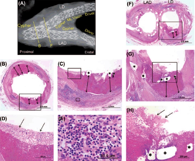
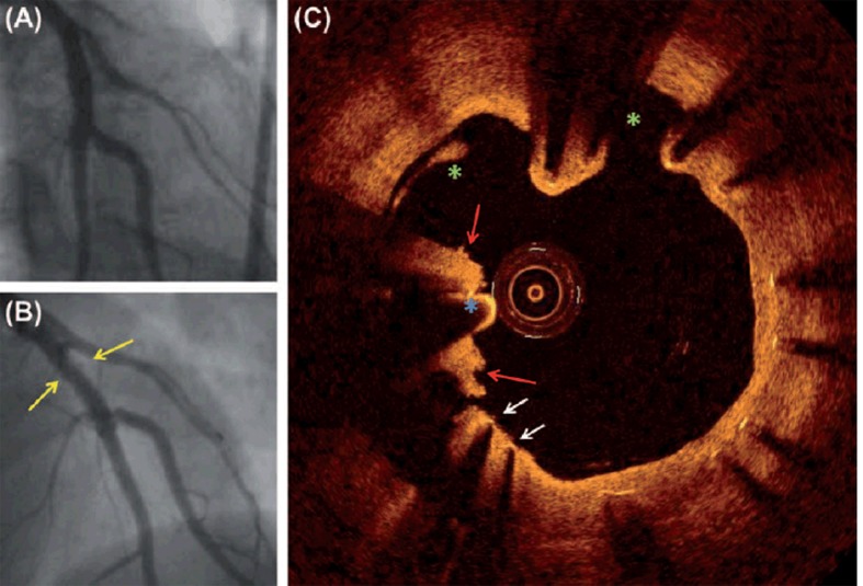
Results: The first patient had ACS 15 months after DES implantation. The angiogram (CAG) was near normal with slight peri-stent contrast staining. OCT revealed abnormalities including thrombus not visible on CAG. These are findings that may explain the ACS. The second patient had subclinical episodes with chest pain after DES implantation. The patient died from stent thrombosis in a DES. Postmortem histological examination of the coronary arteries revealed stent struts with little or no neointimal coverage, persistent peri-strut fibrin deposition, inflammatory cells, malapposition, and small luminal platelet-rich thrombi. Old spotty myocardial infarctions were found in the supplied territory possibly caused by earlier episodes of embolizing thrombus.
Conclusions: In patients with previous implanted DES presenting with ACS, OCT may detect abnormalities and thrombus formation not visible on CAG. Such findings may impact the treatment strategy in these patients.

 求助内容:
求助内容: 应助结果提醒方式:


