H Trimarchi, A Karl, M S Raña, M Forrester, V Pomeranz, F Lombi, A Iotti
{"title":"Initially Nondiagnosed Fabry's Disease when Electron Microscopy Is Lacking: The Continuing Story of Focal and Segmental Glomerulosclerosis.","authors":"H Trimarchi, A Karl, M S Raña, M Forrester, V Pomeranz, F Lombi, A Iotti","doi":"10.1159/000351516","DOIUrl":null,"url":null,"abstract":"<p><p>Focal and segmental glomerulosclerosis is classified as either primary or secondary. We present a patient with a past history of biopsy-proven focal and segmental glomerulosclerosis. Despite initial response to dual blockade and steroids, proteinuria raised when steroids were decreased. After the patient was restarted on steroids, proteinuria did not improve. Another biopsy confirmed the previous diagnosis but suggested Fabry's disease, later confirmed by electron microscopy, α-galactosidase A serum and leukocyte deficiency as well as genetic studies. Proteinuria decreased when agalsidase β was prescribed in parallel with steroid tapering, increased with steroid discontinuation and improved with meprednisone administration. This report highlights the relevance of electron microscopy in kidney biopsy. In glomerulosclerosis, despite specific treatment, secondary hemodynamic and immunologic pathways may contribute to the development of proteinuria and accelerate the renal disease progression due to the primary disease. We discuss possible pathophysiologic pathways involved in proteinuria in Fabry's disease according to the biopsy and the therapeutic response. </p>","PeriodicalId":89663,"journal":{"name":"Case reports in nephrology and urology","volume":" ","pages":"51-7"},"PeriodicalIF":0.0000,"publicationDate":"2013-05-04","publicationTypes":"Journal Article","fieldsOfStudy":null,"isOpenAccess":false,"openAccessPdf":"https://sci-hub-pdf.com/10.1159/000351516","citationCount":"13","resultStr":null,"platform":"Semanticscholar","paperid":null,"PeriodicalName":"Case reports in nephrology and urology","FirstCategoryId":"1085","ListUrlMain":"https://doi.org/10.1159/000351516","RegionNum":0,"RegionCategory":null,"ArticlePicture":[],"TitleCN":null,"AbstractTextCN":null,"PMCID":null,"EPubDate":"2013/1/1 0:00:00","PubModel":"eCollection","JCR":"","JCRName":"","Score":null,"Total":0}
引用次数: 13
Abstract
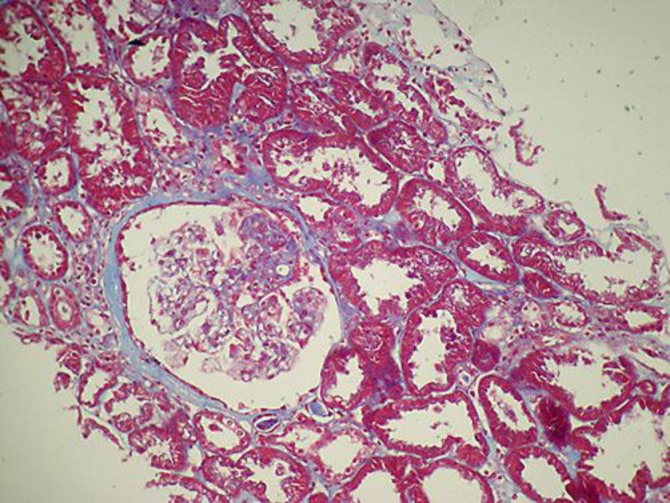
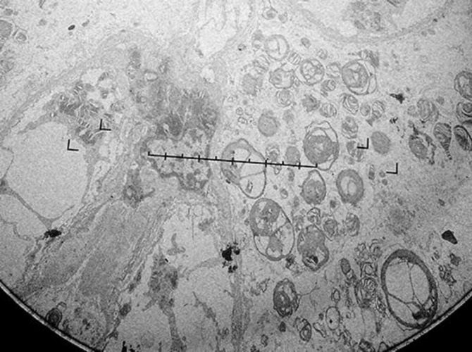
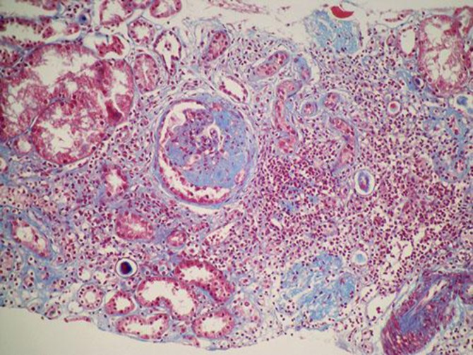
Focal and segmental glomerulosclerosis is classified as either primary or secondary. We present a patient with a past history of biopsy-proven focal and segmental glomerulosclerosis. Despite initial response to dual blockade and steroids, proteinuria raised when steroids were decreased. After the patient was restarted on steroids, proteinuria did not improve. Another biopsy confirmed the previous diagnosis but suggested Fabry's disease, later confirmed by electron microscopy, α-galactosidase A serum and leukocyte deficiency as well as genetic studies. Proteinuria decreased when agalsidase β was prescribed in parallel with steroid tapering, increased with steroid discontinuation and improved with meprednisone administration. This report highlights the relevance of electron microscopy in kidney biopsy. In glomerulosclerosis, despite specific treatment, secondary hemodynamic and immunologic pathways may contribute to the development of proteinuria and accelerate the renal disease progression due to the primary disease. We discuss possible pathophysiologic pathways involved in proteinuria in Fabry's disease according to the biopsy and the therapeutic response.

 求助内容:
求助内容: 应助结果提醒方式:


