Dual fibular allograft dowel technique for sacroiliac joint arthrodesis.
引用次数: 28
Abstract
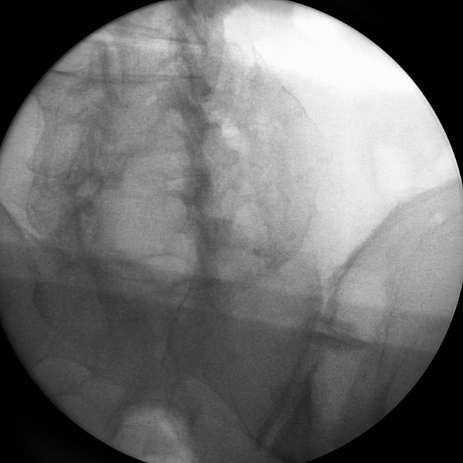
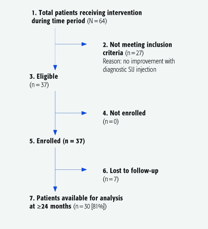
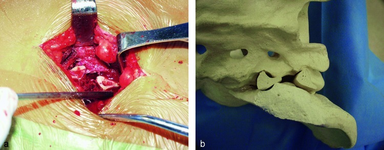
ABSTRACT Study design: Retrospective case series. Objective: To assess fusion rates in patients with sacroiliac joint (SIJ) pain following a minimally invasive technique using fibular dowel allograft. Methods: Thirty-seven consecutive patients (mean age: 42.5 years [range, 23–63 years]) with SIJ pain treated with 38 minimally invasive elective SIJ arthrodeses were retrospectively reviewed using chart and x-ray data. The fusion procedure consisted of minimal muscle stripping over the posterior SIJ and insertion of a cranial and caudal fibular dowel graft across the joint following placement of Steinmann pins. Fusion was deemed to be present when bone bridging trabeculae could be seen crossing the SIJ on either oblique x-rays or by computed tomographic scan. Patients were followed-up for a mean of 52 months (range, 24–62 months). Visual Analog Scale (VAS) was used to monitor clinical pain improvement. Results: Thirty-four patients with SIJ arthrodeses (89.5%) healed and led to substantial improvement in VAS pain scores (preoperative 9.1, postoperative 3.4) (P < .001). This improvement in VAS occurred over a 6-month period and was sustained through subsequent follow-up. Nonunion occurred in four patients with SIJ (10.5%). Each SIJ nonunion was successfully treated by secondary autogenous bone grafting and compression screw fixation. Conclusions: In patients with primary low back pain attributable to the SIJ, a minimally invasive, dual fibular dowel graft provided high rates of fusion and improved pain scores.
双腓骨异体钉技术在骶髂关节融合术中的应用。
研究设计:回顾性病例系列。目的:评估骶髂关节(SIJ)疼痛患者采用同种异体腓骨钉微创技术后的融合率。方法:对连续37例(平均年龄42.5岁[范围23-63岁])行38例微创选择性SIJ关节融合术治疗SIJ疼痛的患者进行回顾性分析,采用图表和x线资料。融合术包括在SIJ后部进行最小程度的肌肉剥离,并在放置Steinmann钉后穿过关节插入颅侧和尾侧腓骨钉移植物。当斜位x线或计算机断层扫描显示骨桥小梁穿过SIJ时,认为存在融合。患者平均随访52个月(24-62个月)。采用视觉模拟评分法(VAS)监测临床疼痛改善情况。结果:34例SIJ关节病患者(89.5%)愈合,VAS疼痛评分显著改善(术前9.1分,术后3.4分)(P)。结论:对于由SIJ引起的原发性腰痛患者,微创双腓骨钉移植物融合率高,疼痛评分改善。[表:见正文]。
本文章由计算机程序翻译,如有差异,请以英文原文为准。
求助全文
约1分钟内获得全文
求助全文

 求助内容:
求助内容: 应助结果提醒方式:


