{"title":"Ultrasound diagnosis of cephalopagus conjoined twin pregnancy at 29 weeks.","authors":"D Sabih, E Ahmad, A Sabih, Q Sabih","doi":"10.2349/biij.6.4.e38","DOIUrl":null,"url":null,"abstract":"<p><p>The authors report a case of a cephalopagus conjoined twin that was diagnosed at 29 weeks of gestation despite the mother having had two ultrasounds done previously. The fetus had one head and face, fused thoraces, common umbilicus but had two pelvises and two sets of genitalia. The fetus had four normally formed legs and arms.Antenatal ultrasound images are supplemented by post natal photographs. A review of literature, clues to ultrasound diagnosis and possible causes of missing this significant abnormality until the 3rd trimester are discussed.</p>","PeriodicalId":89331,"journal":{"name":"Biomedical imaging and intervention journal","volume":"6 4","pages":"e38"},"PeriodicalIF":0.0000,"publicationDate":"2010-10-01","publicationTypes":"Journal Article","fieldsOfStudy":null,"isOpenAccess":false,"openAccessPdf":"https://www.ncbi.nlm.nih.gov/pmc/articles/PMC3097803/pdf/","citationCount":"0","resultStr":null,"platform":"Semanticscholar","paperid":null,"PeriodicalName":"Biomedical imaging and intervention journal","FirstCategoryId":"1085","ListUrlMain":"https://doi.org/10.2349/biij.6.4.e38","RegionNum":0,"RegionCategory":null,"ArticlePicture":[],"TitleCN":null,"AbstractTextCN":null,"PMCID":null,"EPubDate":"","PubModel":"","JCR":"","JCRName":"","Score":null,"Total":0}
引用次数: 0
Abstract
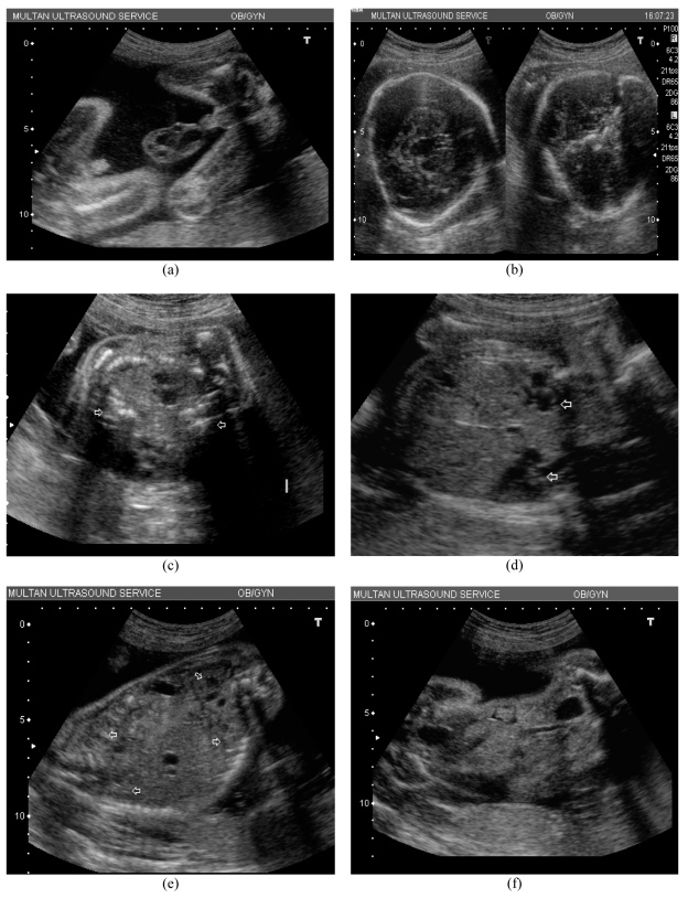
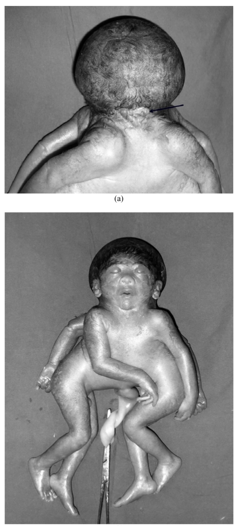
The authors report a case of a cephalopagus conjoined twin that was diagnosed at 29 weeks of gestation despite the mother having had two ultrasounds done previously. The fetus had one head and face, fused thoraces, common umbilicus but had two pelvises and two sets of genitalia. The fetus had four normally formed legs and arms.Antenatal ultrasound images are supplemented by post natal photographs. A review of literature, clues to ultrasound diagnosis and possible causes of missing this significant abnormality until the 3rd trimester are discussed.

 求助内容:
求助内容: 应助结果提醒方式:


