{"title":"Multislice CT angiography of the plantar arch.","authors":"L Field, Z Sun","doi":"10.2349/biij.6.1.e10","DOIUrl":null,"url":null,"abstract":"<p><p>The aim of this case report is to present a multislice computed tomography angiography (CTA) procedure for viewing the plantar arch. A CTA was requested to determine the vascular sufficiency of the plantar arch of a 64-year-old patient with necrotic and gangrenous toes. The patient had recently undergone a proximal wedge osteotomy procedure for correction of a hallux valgus deformity. A 16-detector row CT scanner with 1.25 mm slice thickness and 0.625 mm reconstruction interval was used to reconstruct multiplanar reformats, maximum intensity projections and three-dimensional volume rendered images of the foot in question in both arterial and venous phases to determine if pathology of the plantar arch was present. The 3D reconstructed images of CTA demonstrated a loss of continuity of the plantar arch between the first and third metatarsals. This case report shows the diagnostic value of multislice CTA, especially 3D visualisation in the assessment of peripheral vascular branches.</p>","PeriodicalId":89331,"journal":{"name":"Biomedical imaging and intervention journal","volume":"6 1","pages":"e10"},"PeriodicalIF":0.0000,"publicationDate":"2010-01-01","publicationTypes":"Journal Article","fieldsOfStudy":null,"isOpenAccess":false,"openAccessPdf":"https://sci-hub-pdf.com/10.2349/biij.6.1.e10","citationCount":"0","resultStr":null,"platform":"Semanticscholar","paperid":null,"PeriodicalName":"Biomedical imaging and intervention journal","FirstCategoryId":"1085","ListUrlMain":"https://doi.org/10.2349/biij.6.1.e10","RegionNum":0,"RegionCategory":null,"ArticlePicture":[],"TitleCN":null,"AbstractTextCN":null,"PMCID":null,"EPubDate":"","PubModel":"","JCR":"","JCRName":"","Score":null,"Total":0}
引用次数: 0
Abstract
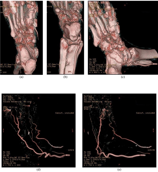
The aim of this case report is to present a multislice computed tomography angiography (CTA) procedure for viewing the plantar arch. A CTA was requested to determine the vascular sufficiency of the plantar arch of a 64-year-old patient with necrotic and gangrenous toes. The patient had recently undergone a proximal wedge osteotomy procedure for correction of a hallux valgus deformity. A 16-detector row CT scanner with 1.25 mm slice thickness and 0.625 mm reconstruction interval was used to reconstruct multiplanar reformats, maximum intensity projections and three-dimensional volume rendered images of the foot in question in both arterial and venous phases to determine if pathology of the plantar arch was present. The 3D reconstructed images of CTA demonstrated a loss of continuity of the plantar arch between the first and third metatarsals. This case report shows the diagnostic value of multislice CTA, especially 3D visualisation in the assessment of peripheral vascular branches.

 求助内容:
求助内容: 应助结果提醒方式:


