{"title":"A new method for experimental characterisation of scattered radiation in 64-slice CT scanner.","authors":"A Akbarzadeh, Mr Ay, H Ghadiri, S Sarkar, H Zaidi","doi":"10.2349/biij.6.1.e3","DOIUrl":null,"url":null,"abstract":"<p><strong>Purpose: </strong>The consummate 64-slice CT scanner that spawns a new generation of non-invasive diagnostic tool, however revolutionary, brings with it the incidental by-product that is scattered radiation. The extended detector aperture capability in the 64-slcie CT scanner allows the effects of scattered radiation to be more pronounced and therefore demands that the magnitude and spatial distribution of scatter component be addressed during the imaging process. To this end, corrective algorithms need to be formulated on a basis of a precise understanding of scatter distribution. Relative to a 64-slice CT scanner, here now a unique solution is based upon dedicated blockers operative within various detector rows, calculating scatter profiles and scatter to primary ratios (SPR).</p><p><strong>Materials and methods: </strong>A single dimension blocker array was installed beneath the collimator, and the extrapolated shadow area on the detectors revealed the scatter radiation after exposure. The experiment was conducted using a 64-slice CT scanner manufactured by GE Healthcare Technologies.</p><p><strong>Results: </strong>Variables such as tube voltage, phantom size and phantom-off centring on the scatter profile and the SPR was measured using the dedicated blocker method introduced above. When tube voltage is increased from 80kVp to 140kVp in a 21.5 cm water phantom, the SPR is found to reduce from 219.9 to 39.9 respectively.</p><p><strong>Conclusion: </strong>The method developed within this study is applicable to any measurement and is direct with minimal complexity.</p>","PeriodicalId":89331,"journal":{"name":"Biomedical imaging and intervention journal","volume":"6 1","pages":"e3"},"PeriodicalIF":0.0000,"publicationDate":"2010-01-01","publicationTypes":"Journal Article","fieldsOfStudy":null,"isOpenAccess":false,"openAccessPdf":"https://sci-hub-pdf.com/10.2349/biij.6.1.e3","citationCount":"2","resultStr":null,"platform":"Semanticscholar","paperid":null,"PeriodicalName":"Biomedical imaging and intervention journal","FirstCategoryId":"1085","ListUrlMain":"https://doi.org/10.2349/biij.6.1.e3","RegionNum":0,"RegionCategory":null,"ArticlePicture":[],"TitleCN":null,"AbstractTextCN":null,"PMCID":null,"EPubDate":"","PubModel":"","JCR":"","JCRName":"","Score":null,"Total":0}
引用次数: 2
Abstract
Purpose: The consummate 64-slice CT scanner that spawns a new generation of non-invasive diagnostic tool, however revolutionary, brings with it the incidental by-product that is scattered radiation. The extended detector aperture capability in the 64-slcie CT scanner allows the effects of scattered radiation to be more pronounced and therefore demands that the magnitude and spatial distribution of scatter component be addressed during the imaging process. To this end, corrective algorithms need to be formulated on a basis of a precise understanding of scatter distribution. Relative to a 64-slice CT scanner, here now a unique solution is based upon dedicated blockers operative within various detector rows, calculating scatter profiles and scatter to primary ratios (SPR).
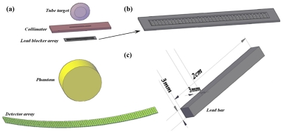
Materials and methods: A single dimension blocker array was installed beneath the collimator, and the extrapolated shadow area on the detectors revealed the scatter radiation after exposure. The experiment was conducted using a 64-slice CT scanner manufactured by GE Healthcare Technologies.
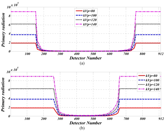
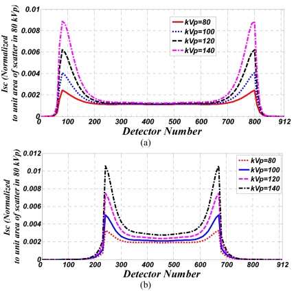
Results: Variables such as tube voltage, phantom size and phantom-off centring on the scatter profile and the SPR was measured using the dedicated blocker method introduced above. When tube voltage is increased from 80kVp to 140kVp in a 21.5 cm water phantom, the SPR is found to reduce from 219.9 to 39.9 respectively.
Conclusion: The method developed within this study is applicable to any measurement and is direct with minimal complexity.

 求助内容:
求助内容: 应助结果提醒方式:


