{"title":"An intense F-FDG pulmonary microfocus on PET without detectable abnormality on CT: A manifestation of an iatrogenic FDG pulmonary embolus.","authors":"As Fathinul Fikri, Wfe Lau","doi":"10.2349/biij.6.4.e37","DOIUrl":null,"url":null,"abstract":"<p><p>An incidental finding of an intense focus of (18)F-Fluorodeoxyglucose (FDG) pulmonary uptake on positron emission tomography (PET) without detectable lesions on computed tomography (CT) is highly suggestive of FDG microembolus. Its microscopic nature means it is undetectable on CT. It is an artefact attributable to (18)F-FDG-tracer contamination at the injection site. This paper reports a case of a 61 year-old lady with a past history of breast carcinoma, in whom follow-up PET/CT images demonstrated an incidental intense FDG pulmonary abnormality. A follow-up PET/CT seven months later demonstrated complete resolution of the abnormality.</p>","PeriodicalId":89331,"journal":{"name":"Biomedical imaging and intervention journal","volume":"6 4","pages":"e37"},"PeriodicalIF":0.0000,"publicationDate":"2010-10-01","publicationTypes":"Journal Article","fieldsOfStudy":null,"isOpenAccess":false,"openAccessPdf":"https://www.ncbi.nlm.nih.gov/pmc/articles/PMC3097806/pdf/","citationCount":"12","resultStr":null,"platform":"Semanticscholar","paperid":null,"PeriodicalName":"Biomedical imaging and intervention journal","FirstCategoryId":"1085","ListUrlMain":"https://doi.org/10.2349/biij.6.4.e37","RegionNum":0,"RegionCategory":null,"ArticlePicture":[],"TitleCN":null,"AbstractTextCN":null,"PMCID":null,"EPubDate":"","PubModel":"","JCR":"","JCRName":"","Score":null,"Total":0}
引用次数: 12
Abstract
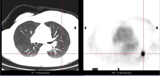
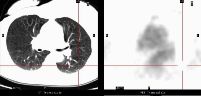
An incidental finding of an intense focus of (18)F-Fluorodeoxyglucose (FDG) pulmonary uptake on positron emission tomography (PET) without detectable lesions on computed tomography (CT) is highly suggestive of FDG microembolus. Its microscopic nature means it is undetectable on CT. It is an artefact attributable to (18)F-FDG-tracer contamination at the injection site. This paper reports a case of a 61 year-old lady with a past history of breast carcinoma, in whom follow-up PET/CT images demonstrated an incidental intense FDG pulmonary abnormality. A follow-up PET/CT seven months later demonstrated complete resolution of the abnormality.

 求助内容:
求助内容: 应助结果提醒方式:


