{"title":"F-FDG PET/CT as a potential valuable adjunct to MRI in characterising the Brodie's abscess.","authors":"F Fathinul, Aj Nordin","doi":"10.2349/biij.6.3.e26","DOIUrl":null,"url":null,"abstract":"<p><p>Chronic osteomyelitis (Brodie's abscess) is essentially a problem of diagnosis, and there may be considerable difficulty in distinguishing it from other benign and malignant bone lesions. Early diagnosis of Brodie's abscess is deemed important as the disease has a good curative potential following an appropriate antibiotic treatment. Of late, PET/CT using (18)F-FDG is taking a centre stage in the imaging of bone infection though documentation on its role in characterising the feature of Brodie's abscess is exceedingly scarce. On the other hand, it is well known that MRI imaging plays a very important role in distinguishing abscess loculation from malignancy. The authors present the case of a 13-year-old boy with pain in the right heel for few months. Radiograph of the right foot revealed a lucent focus with sclerotic margin in the right calcaneum. MRI T1-weighted images were inconclusive of penumbra sign to characterise abscess cavity due to the small volume lesion. Whole-body (18)F-FDG PET/CT scan showed multiple small avid lesions at the margin of the sclerotic rim in the right calcaneum. Final diagnosis of Brodie's abscess with Klebsiella culture was confirmed via bone debridement.</p>","PeriodicalId":89331,"journal":{"name":"Biomedical imaging and intervention journal","volume":"6 3","pages":"e26"},"PeriodicalIF":0.0000,"publicationDate":"2010-07-01","publicationTypes":"Journal Article","fieldsOfStudy":null,"isOpenAccess":false,"openAccessPdf":"https://sci-hub-pdf.com/10.2349/biij.6.3.e26","citationCount":"7","resultStr":null,"platform":"Semanticscholar","paperid":null,"PeriodicalName":"Biomedical imaging and intervention journal","FirstCategoryId":"1085","ListUrlMain":"https://doi.org/10.2349/biij.6.3.e26","RegionNum":0,"RegionCategory":null,"ArticlePicture":[],"TitleCN":null,"AbstractTextCN":null,"PMCID":null,"EPubDate":"","PubModel":"","JCR":"","JCRName":"","Score":null,"Total":0}
引用次数: 7
Abstract
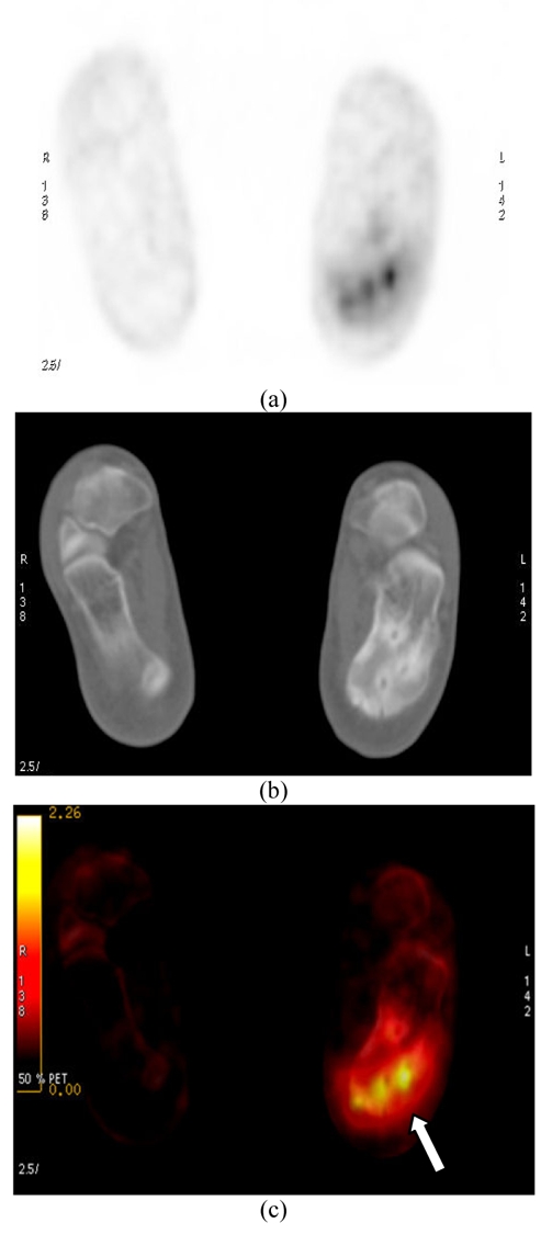
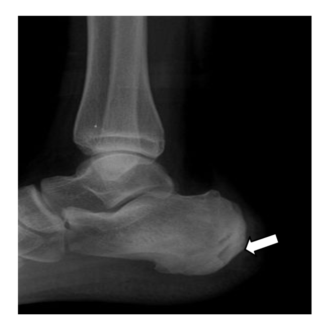
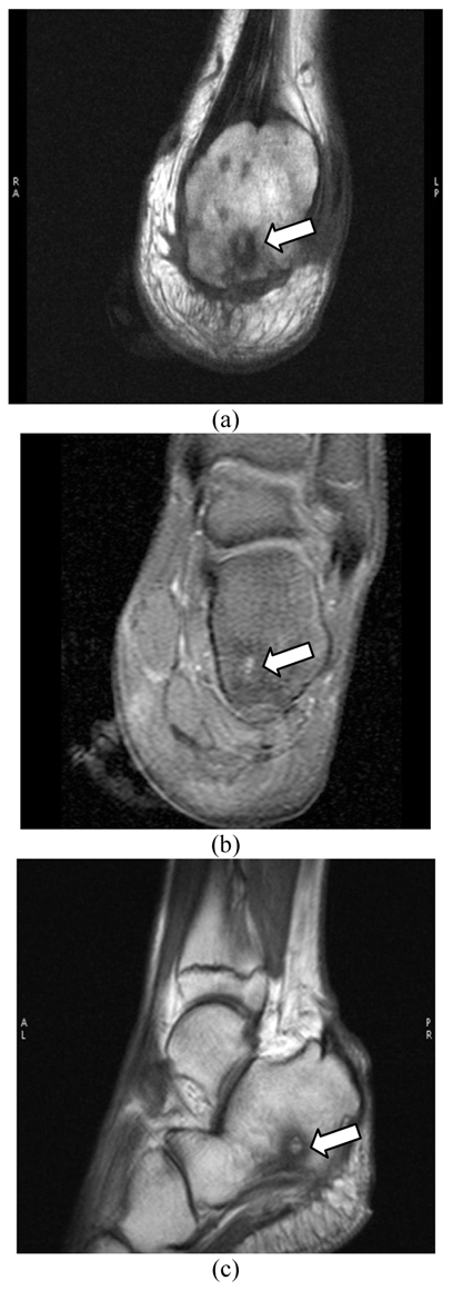
Chronic osteomyelitis (Brodie's abscess) is essentially a problem of diagnosis, and there may be considerable difficulty in distinguishing it from other benign and malignant bone lesions. Early diagnosis of Brodie's abscess is deemed important as the disease has a good curative potential following an appropriate antibiotic treatment. Of late, PET/CT using (18)F-FDG is taking a centre stage in the imaging of bone infection though documentation on its role in characterising the feature of Brodie's abscess is exceedingly scarce. On the other hand, it is well known that MRI imaging plays a very important role in distinguishing abscess loculation from malignancy. The authors present the case of a 13-year-old boy with pain in the right heel for few months. Radiograph of the right foot revealed a lucent focus with sclerotic margin in the right calcaneum. MRI T1-weighted images were inconclusive of penumbra sign to characterise abscess cavity due to the small volume lesion. Whole-body (18)F-FDG PET/CT scan showed multiple small avid lesions at the margin of the sclerotic rim in the right calcaneum. Final diagnosis of Brodie's abscess with Klebsiella culture was confirmed via bone debridement.

 求助内容:
求助内容: 应助结果提醒方式:


