Haruhisa Okawa, Johan Pahlberg, Fred Rieke, Lutz Birnbaumer, Alapakkam P Sampath
{"title":"Coordinated control of sensitivity by two splice variants of Gα(o) in retinal ON bipolar cells.","authors":"Haruhisa Okawa, Johan Pahlberg, Fred Rieke, Lutz Birnbaumer, Alapakkam P Sampath","doi":"10.1085/jgp.201010477","DOIUrl":null,"url":null,"abstract":"<p><p>The high sensitivity of scotopic vision depends on the efficient retinal processing of single photon responses generated by individual rod photoreceptors. At the first synapse in the mammalian retina, rod outputs are pooled by a rod \"ON\" bipolar cell, which uses a G-protein signaling cascade to enhance the fidelity of the single photon response under conditions where few rods absorb light. Here we show in mouse rod bipolar cells that both splice variants of the G(o) α subunit, Gα(o1) and Gα(o2), mediate light responses under the control of mGluR6 receptors, and their coordinated action is critical for maximizing sensitivity. We found that the light response of rod bipolar cells was primarily mediated by Gα(o1), but the loss of Gα(o2) caused a reduction in the light sensitivity. This reduced sensitivity was not attributable to the reduction in the total number of G(o) α subunits, or the altered balance of expression levels between the two splice variants. These results indicate that Gα(o1) and Gα(o2) both mediate a depolarizing light response in rod bipolar cells without occluding each other's actions, suggesting they might act independently on a common effector. Thus, Gα(o2) plays a role in improving the sensitivity of rod bipolar cells through its action with Gα(o1). The coordinated action of two splice variants of a single Gα may represent a novel mechanism for the fine control of G-protein activity.</p>","PeriodicalId":173753,"journal":{"name":"The Journal of General Physiology","volume":" ","pages":"443-54"},"PeriodicalIF":0.0000,"publicationDate":"2010-10-01","publicationTypes":"Journal Article","fieldsOfStudy":null,"isOpenAccess":false,"openAccessPdf":"https://ftp.ncbi.nlm.nih.gov/pub/pmc/oa_pdf/d6/cd/JGP_201010477.PMC2947061.pdf","citationCount":"0","resultStr":null,"platform":"Semanticscholar","paperid":null,"PeriodicalName":"The Journal of General Physiology","FirstCategoryId":"1085","ListUrlMain":"https://doi.org/10.1085/jgp.201010477","RegionNum":0,"RegionCategory":null,"ArticlePicture":[],"TitleCN":null,"AbstractTextCN":null,"PMCID":null,"EPubDate":"2010/9/13 0:00:00","PubModel":"Epub","JCR":"","JCRName":"","Score":null,"Total":0}
引用次数: 0
Abstract
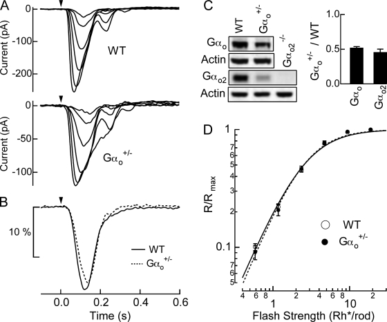
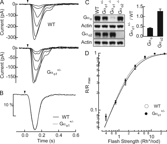
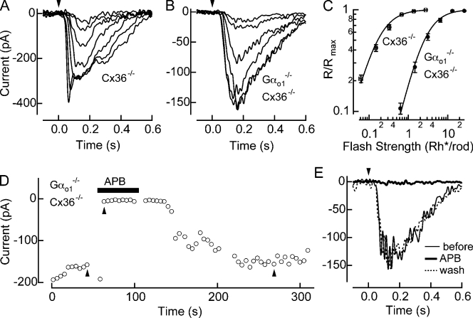
The high sensitivity of scotopic vision depends on the efficient retinal processing of single photon responses generated by individual rod photoreceptors. At the first synapse in the mammalian retina, rod outputs are pooled by a rod "ON" bipolar cell, which uses a G-protein signaling cascade to enhance the fidelity of the single photon response under conditions where few rods absorb light. Here we show in mouse rod bipolar cells that both splice variants of the G(o) α subunit, Gα(o1) and Gα(o2), mediate light responses under the control of mGluR6 receptors, and their coordinated action is critical for maximizing sensitivity. We found that the light response of rod bipolar cells was primarily mediated by Gα(o1), but the loss of Gα(o2) caused a reduction in the light sensitivity. This reduced sensitivity was not attributable to the reduction in the total number of G(o) α subunits, or the altered balance of expression levels between the two splice variants. These results indicate that Gα(o1) and Gα(o2) both mediate a depolarizing light response in rod bipolar cells without occluding each other's actions, suggesting they might act independently on a common effector. Thus, Gα(o2) plays a role in improving the sensitivity of rod bipolar cells through its action with Gα(o1). The coordinated action of two splice variants of a single Gα may represent a novel mechanism for the fine control of G-protein activity.

 求助内容:
求助内容: 应助结果提醒方式:


