Identification of cysteine proteases and screening of cysteine protease inhibitors in biological samples by a two-dimensional gel system of zymography and reverse zymography.
{"title":"Identification of cysteine proteases and screening of cysteine protease inhibitors in biological samples by a two-dimensional gel system of zymography and reverse zymography.","authors":"Eiichi Saitoh, Shinya Yamamoto, Eishiro Okamoto, Yoshimi Hayakawa, Takashi Hoshino, Ritsuko Sato, Satoko Isemura, Sadami Ohtsubo, Masayuki Taniguchi","doi":"","DOIUrl":null,"url":null,"abstract":"<p><p>We have developed a two-dimensional (2D-) gel system of zymography and reverse zymography for the detection and characterization of proteases and protease inhibitors. Isoelectric focusing (IEF) agarose gels with pH gradients were employed for separation in the first-dimension and sodium dodecyl sulfate (SDS)-polyacrylamide gel copolymerized with gelatin used for the second dimension. Proteases and protease inhibitors separated by IEF gel were applied on the second gel without trichloroacetic acid (TCA) fixation. Protease activity in the 2D-gel was visualized as transparent spots where gelatin substrate was digested after commassie brilliant blue (CBB) staining. Some of the transparent spots from the skin mucus extract of rainbow trout were determined to be a cysteine protease through use of E-64 or CA-074. In the reverse zymography technique, the gel was incubated with papain solution at 37 degrees C for 18 h. Cysteine protease inhibitors from broad bean seeds were detected as clear blue spots after CBB staining. The amino (N-) terminal sequences of four papain inhibitor spots thus detected were demonstrated to be identical to that of favin beta chain, a broad bean lectin. Taken together, our system can be considered to be an efficient technique for discovering and characterizing new proteases and protease inhibitors in biological samples. This is the first report describing a 2D-gel system of zymography and reverse zymography.</p>","PeriodicalId":7781,"journal":{"name":"Analytical Chemistry Insights","volume":"2 ","pages":"51-9"},"PeriodicalIF":0.0000,"publicationDate":"2007-11-18","publicationTypes":"Journal Article","fieldsOfStudy":null,"isOpenAccess":false,"openAccessPdf":"https://www.ncbi.nlm.nih.gov/pmc/articles/PMC2716818/pdf/","citationCount":"0","resultStr":null,"platform":"Semanticscholar","paperid":null,"PeriodicalName":"Analytical Chemistry Insights","FirstCategoryId":"1085","ListUrlMain":"","RegionNum":0,"RegionCategory":null,"ArticlePicture":[],"TitleCN":null,"AbstractTextCN":null,"PMCID":null,"EPubDate":"","PubModel":"","JCR":"","JCRName":"","Score":null,"Total":0}
引用次数: 0
Abstract
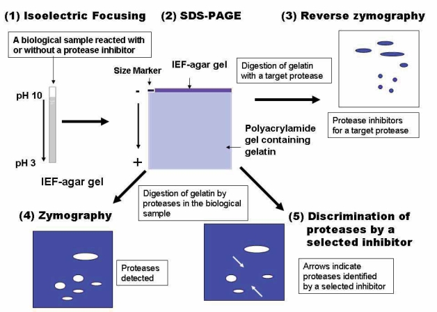
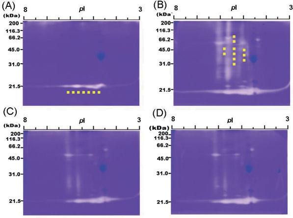
We have developed a two-dimensional (2D-) gel system of zymography and reverse zymography for the detection and characterization of proteases and protease inhibitors. Isoelectric focusing (IEF) agarose gels with pH gradients were employed for separation in the first-dimension and sodium dodecyl sulfate (SDS)-polyacrylamide gel copolymerized with gelatin used for the second dimension. Proteases and protease inhibitors separated by IEF gel were applied on the second gel without trichloroacetic acid (TCA) fixation. Protease activity in the 2D-gel was visualized as transparent spots where gelatin substrate was digested after commassie brilliant blue (CBB) staining. Some of the transparent spots from the skin mucus extract of rainbow trout were determined to be a cysteine protease through use of E-64 or CA-074. In the reverse zymography technique, the gel was incubated with papain solution at 37 degrees C for 18 h. Cysteine protease inhibitors from broad bean seeds were detected as clear blue spots after CBB staining. The amino (N-) terminal sequences of four papain inhibitor spots thus detected were demonstrated to be identical to that of favin beta chain, a broad bean lectin. Taken together, our system can be considered to be an efficient technique for discovering and characterizing new proteases and protease inhibitors in biological samples. This is the first report describing a 2D-gel system of zymography and reverse zymography.


 求助内容:
求助内容: 应助结果提醒方式:


