Jin Joo Choi, Dong Woo Park, Dong Hyun Ahn, Woo-Suk Tae, Jin-Hwa Moon
{"title":"Decreased Brain Surface Complexity in Children With Attention Deficit Hyperactivity Disorder.","authors":"Jin Joo Choi, Dong Woo Park, Dong Hyun Ahn, Woo-Suk Tae, Jin-Hwa Moon","doi":"10.3988/jcn.2022.18.1.123","DOIUrl":null,"url":null,"abstract":"Dear Editor, Various neuroimaging studies of attention deficit hyperactivity disorder (ADHD) have revealed abnormalities in the frontal cortex, left basal ganglia, and left inferior frontal gyrus.1 However, the results of these studies have been inconsistent and usually not replicable.1,2 To date, there is not a single neuroimaging marker for ADHD diagnosis. Surface-based morphometry (SBM) can be used to analyze various characteristics of the brain surface and estimate the cortical thickness (CT), local gyrification index (LGI), surface area, and fractal dimension (FD). Previous large-scale studies of CT revealed subtle differences between ADHD children and controls,1 whereas the few studies of LGI have found no difference between ADHD and control groups. FD characterizes the fractal patterns of geometric objects, and is currently used to estimate the cortical complexity. However, very few studies have applied FD analysis to ADHD.3 This study aimed to compare various SBM characteristics between ADHD children and controls. Children aged 8–15 years who were diagnosed with ADHD and age-matched controls who voluntarily participated were included. Patients with structural brain lesions, severe medical problems, full-scale intelligence quotient (FSIQ) <80, or impairments in hearing, vision, or movement were excluded. Neurological and neuropsychological examinations and neurocognitive function tests (NCFTs) were performed. ADHD was diagnosed based on ADHD Rating Scale-IV. Magnetic resonance imaging (MRI) was performed using a 3-T MRI scanner. The detailed acquisition methods are presented in Supplementary Table 1 (in the onlineonly Data Supplement). For SBM, coronal three-dimensional T1-weighted structural images were processed using the Computational Anatomy Toolbox (version 12, http://www.neuro. uni-jena.de/cat/). Group differences in CT, LGI, and FD were tested separately using analysis of covariance with age and sex as with 5,000 permutations. The multiple-comparison problem was corrected at the cluster level using threshold-free cluster enhancement to a family-wise error (TFCE-FWE), with a rate of p<0.05. The study included 11 patients and 19 controls, whose detailed demographics and NCFT data are presented in Supplementary Table 2 (in the online-only Data Supplement). Age, righthandedness, male-to-female ratio, and FSIQ did not differ significantly between the groups. The SBM analysis did not reveal any significant differences in CT and LGI between the groups. However, FD analysis showed significant group differences in multiple regions, with FD being lower in ADHD patients than in controls. Although group differences were observed in both the right and left cortexes, FD of the ADHD group was decreased most significantly (TFCEFWE-corrected p<0.05) in the left frontal, parietal, cingulate, and occipital cortexes (Fig. 1). No region in the ADHD group showed an increased FD. This study found that the cortical FD is significantly decreased in ADHD children, especially in the left cortex. A decreased FD in ADHD indicates an altered geometric complexity of the brain surface, which implicates low efficiency of the corresponding brain regions in perJin Joo Choi Dong Woo Park Dong Hyun Ahn Woo-Suk Tae Jin-Hwa Moon","PeriodicalId":324902,"journal":{"name":"Journal of Clinical Neurology (Seoul, Korea)","volume":" ","pages":"123-125"},"PeriodicalIF":0.0000,"publicationDate":"2022-01-01","publicationTypes":"Journal Article","fieldsOfStudy":null,"isOpenAccess":false,"openAccessPdf":"https://ftp.ncbi.nlm.nih.gov/pub/pmc/oa_pdf/83/a6/jcn-18-123.PMC8762512.pdf","citationCount":"1","resultStr":null,"platform":"Semanticscholar","paperid":null,"PeriodicalName":"Journal of Clinical Neurology (Seoul, Korea)","FirstCategoryId":"3","ListUrlMain":"https://doi.org/10.3988/jcn.2022.18.1.123","RegionNum":0,"RegionCategory":null,"ArticlePicture":[],"TitleCN":null,"AbstractTextCN":null,"PMCID":null,"EPubDate":"","PubModel":"","JCR":"","JCRName":"","Score":null,"Total":0}
引用次数: 1
Abstract
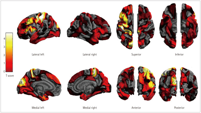
Dear Editor, Various neuroimaging studies of attention deficit hyperactivity disorder (ADHD) have revealed abnormalities in the frontal cortex, left basal ganglia, and left inferior frontal gyrus.1 However, the results of these studies have been inconsistent and usually not replicable.1,2 To date, there is not a single neuroimaging marker for ADHD diagnosis. Surface-based morphometry (SBM) can be used to analyze various characteristics of the brain surface and estimate the cortical thickness (CT), local gyrification index (LGI), surface area, and fractal dimension (FD). Previous large-scale studies of CT revealed subtle differences between ADHD children and controls,1 whereas the few studies of LGI have found no difference between ADHD and control groups. FD characterizes the fractal patterns of geometric objects, and is currently used to estimate the cortical complexity. However, very few studies have applied FD analysis to ADHD.3 This study aimed to compare various SBM characteristics between ADHD children and controls. Children aged 8–15 years who were diagnosed with ADHD and age-matched controls who voluntarily participated were included. Patients with structural brain lesions, severe medical problems, full-scale intelligence quotient (FSIQ) <80, or impairments in hearing, vision, or movement were excluded. Neurological and neuropsychological examinations and neurocognitive function tests (NCFTs) were performed. ADHD was diagnosed based on ADHD Rating Scale-IV. Magnetic resonance imaging (MRI) was performed using a 3-T MRI scanner. The detailed acquisition methods are presented in Supplementary Table 1 (in the onlineonly Data Supplement). For SBM, coronal three-dimensional T1-weighted structural images were processed using the Computational Anatomy Toolbox (version 12, http://www.neuro. uni-jena.de/cat/). Group differences in CT, LGI, and FD were tested separately using analysis of covariance with age and sex as with 5,000 permutations. The multiple-comparison problem was corrected at the cluster level using threshold-free cluster enhancement to a family-wise error (TFCE-FWE), with a rate of p<0.05. The study included 11 patients and 19 controls, whose detailed demographics and NCFT data are presented in Supplementary Table 2 (in the online-only Data Supplement). Age, righthandedness, male-to-female ratio, and FSIQ did not differ significantly between the groups. The SBM analysis did not reveal any significant differences in CT and LGI between the groups. However, FD analysis showed significant group differences in multiple regions, with FD being lower in ADHD patients than in controls. Although group differences were observed in both the right and left cortexes, FD of the ADHD group was decreased most significantly (TFCEFWE-corrected p<0.05) in the left frontal, parietal, cingulate, and occipital cortexes (Fig. 1). No region in the ADHD group showed an increased FD. This study found that the cortical FD is significantly decreased in ADHD children, especially in the left cortex. A decreased FD in ADHD indicates an altered geometric complexity of the brain surface, which implicates low efficiency of the corresponding brain regions in perJin Joo Choi Dong Woo Park Dong Hyun Ahn Woo-Suk Tae Jin-Hwa Moon

 求助内容:
求助内容: 应助结果提醒方式:


