{"title":"Junctophilins 1, 2, and 3 all support voltage-induced Ca2+ release despite considerable divergence.","authors":"Stefano Perni, Kurt Beam","doi":"10.1085/jgp.202113024","DOIUrl":null,"url":null,"abstract":"<p><p>In skeletal muscle, depolarization of the plasma membrane (PM) causes conformational changes of the calcium channel CaV1.1 that then activate RYR1 to release calcium from the SR. Being independent of extracellular calcium entry, this process is termed voltage-induced calcium release. In skeletal muscle, junctophilins (JPHs) 1 and 2 form the SR-PM junctions at which voltage-induced calcium release occurs. Previous work demonstrated that JPH2 is able to recapitulate voltage-induced calcium release when expressed in HEK293 cells together with CaV1.1, β1a, Stac3, and RYR1. However, it is unknown whether JPH1 and the more distantly related neuronal JPH3 and JPH4 might also function in this manner, a question of interest because different JPH isoforms diverge in their interactions with RYR1. Here, we show that, like JPH2, JPH1 and JPH3, coexpressed with CaV1.1, β1a, Stac3, and RYR1 in HEK293 cells, cause colocalization of CaV1.1 and RYR1 at ER-PM junctions. Furthermore, potassium depolarization elicited cytoplasmic calcium transients in cells in which WT CaV1.1 was replaced with the calcium impermeant mutant CaV1.1(N617D), indicating that JPH1, JPH2, and JPH3 can all support voltage-induced calcium release, despite sequence divergence and differences in interaction with RYR1. Conversely, JPH4-induced ER-PM junctions contain CaV1.1 but not RYR1, and cells expressing JPH4 are unable to produce depolarization-induced calcium transients. Thus, JPHs seem to act primarily to form ER-PM junctions and to recruit the necessary signaling proteins to these junctions but appear not to be directly involved in the functional interactions between these proteins.</p>","PeriodicalId":173753,"journal":{"name":"The Journal of General Physiology","volume":" ","pages":""},"PeriodicalIF":0.0000,"publicationDate":"2022-09-05","publicationTypes":"Journal Article","fieldsOfStudy":null,"isOpenAccess":false,"openAccessPdf":"https://ftp.ncbi.nlm.nih.gov/pub/pmc/oa_pdf/c2/e2/JGP_202113024.PMC9488633.pdf","citationCount":"2","resultStr":null,"platform":"Semanticscholar","paperid":null,"PeriodicalName":"The Journal of General Physiology","FirstCategoryId":"3","ListUrlMain":"https://doi.org/10.1085/jgp.202113024","RegionNum":0,"RegionCategory":null,"ArticlePicture":[],"TitleCN":null,"AbstractTextCN":null,"PMCID":null,"EPubDate":"2022/1/28 0:00:00","PubModel":"Epub","JCR":"","JCRName":"","Score":null,"Total":0}
引用次数: 2
Abstract
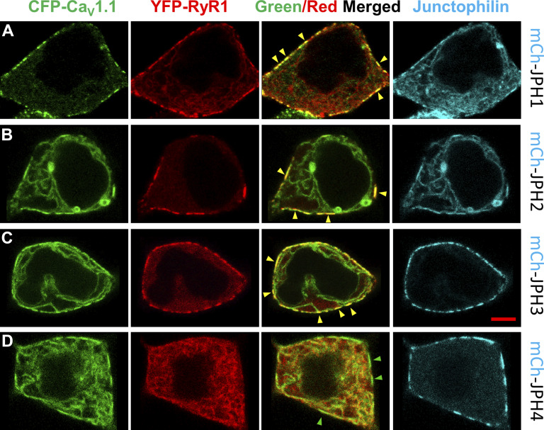
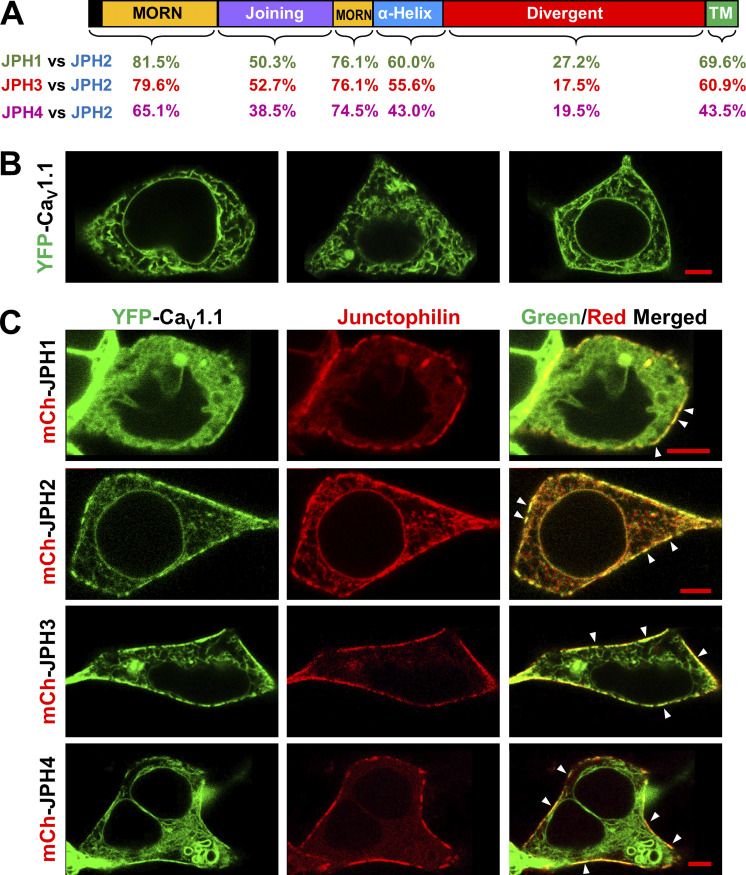
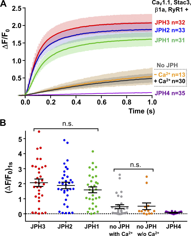
In skeletal muscle, depolarization of the plasma membrane (PM) causes conformational changes of the calcium channel CaV1.1 that then activate RYR1 to release calcium from the SR. Being independent of extracellular calcium entry, this process is termed voltage-induced calcium release. In skeletal muscle, junctophilins (JPHs) 1 and 2 form the SR-PM junctions at which voltage-induced calcium release occurs. Previous work demonstrated that JPH2 is able to recapitulate voltage-induced calcium release when expressed in HEK293 cells together with CaV1.1, β1a, Stac3, and RYR1. However, it is unknown whether JPH1 and the more distantly related neuronal JPH3 and JPH4 might also function in this manner, a question of interest because different JPH isoforms diverge in their interactions with RYR1. Here, we show that, like JPH2, JPH1 and JPH3, coexpressed with CaV1.1, β1a, Stac3, and RYR1 in HEK293 cells, cause colocalization of CaV1.1 and RYR1 at ER-PM junctions. Furthermore, potassium depolarization elicited cytoplasmic calcium transients in cells in which WT CaV1.1 was replaced with the calcium impermeant mutant CaV1.1(N617D), indicating that JPH1, JPH2, and JPH3 can all support voltage-induced calcium release, despite sequence divergence and differences in interaction with RYR1. Conversely, JPH4-induced ER-PM junctions contain CaV1.1 but not RYR1, and cells expressing JPH4 are unable to produce depolarization-induced calcium transients. Thus, JPHs seem to act primarily to form ER-PM junctions and to recruit the necessary signaling proteins to these junctions but appear not to be directly involved in the functional interactions between these proteins.

 求助内容:
求助内容: 应助结果提醒方式:


