Proteomic analysis of extracellular vesicles secreted by primary human epithelial endometrial cells reveals key proteins related to embryo implantation.
Marina Segura-Benítez, María Cristina Carbajo-García, Ana Corachán, Amparo Faus, Antonio Pellicer, Hortensia Ferrero
{"title":"Proteomic analysis of extracellular vesicles secreted by primary human epithelial endometrial cells reveals key proteins related to embryo implantation.","authors":"Marina Segura-Benítez, María Cristina Carbajo-García, Ana Corachán, Amparo Faus, Antonio Pellicer, Hortensia Ferrero","doi":"10.1186/s12958-021-00879-x","DOIUrl":null,"url":null,"abstract":"<p><strong>Background: </strong>Successful implantation is dependent on coordination between maternal endometrium and embryo, and the role of EVs in the required cross-talk cell-to-cell has been recently established. In this regard, it has been reported that EVs secreted by the maternal endometrium can be internalized by human trophoblastic cells transferring their contents and enhancing their adhesive and invasive capacity. This is the first study to comprehensively evaluate three EV isolation methods on human endometrial epithelial cells in culture and to describe the proteomic content of EVs secreted by pHEECs from fertile women.</p><p><strong>Methods: </strong>Ishikawa cells and pHEECs were in vitro cultured and hormonally treated; subsequently, conditioned medium was collected and EVs isolated. Ishikawa cells were used for the comparison of EVs isolation methods ultracentrifugation, ExoQuick-TC and Norgen Cell Culture Media Exosome Purification Kit (n = 3 replicates/isolation method). pHEECs were isolated from endometrial biopsies (n = 8/replicate; 3 replicates) collected from healthy oocyte donors with confirmed fertility, and protein content of EVs isolated by the most efficient methodology was analysed using liquid chromatography-tandem mass spectrometry. EV concentration and size were analyzed by nanoparticle tracking analysis, EV morphology visualized by transmission electron microscopy and protein marker expression was determined by Western blotting.</p><p><strong>Results: </strong>Ultracentrifugation was the most efficient methodology for EV isolation from medium of endometrial epithelial cells. EVs secreted by pHEECs and isolated by ultracentrifugation were heterogeneous in size and expressed EV protein markers HSP70, TSG101, CD9, and CD81. Proteomic analysis identified 218 proteins contained in these EVs enriched in biological processes involved in embryo implantation, including cell adhesion, differentiation, communication, migration, extracellular matrix organization, vasculature development, and reproductive processes. From these proteins, 82 were selected based on their functional relevance in implantation success as possible implantation biomarkers.</p><p><strong>Conclusions: </strong>EV protein cargos are implicated in biological processes related to endometrial receptivity, embryo implantation, and early embryo development, supporting the concept of a communication system between the embryo and the maternal endometrium via EVs. Identified proteins may define new biomarkers of endometrial receptivity and implantation success.</p>","PeriodicalId":520764,"journal":{"name":"Reproductive biology and endocrinology : RB&E","volume":" ","pages":"3"},"PeriodicalIF":0.0000,"publicationDate":"2022-01-03","publicationTypes":"Journal Article","fieldsOfStudy":null,"isOpenAccess":false,"openAccessPdf":"https://www.ncbi.nlm.nih.gov/pmc/articles/PMC8722215/pdf/","citationCount":"20","resultStr":null,"platform":"Semanticscholar","paperid":null,"PeriodicalName":"Reproductive biology and endocrinology : RB&E","FirstCategoryId":"3","ListUrlMain":"https://doi.org/10.1186/s12958-021-00879-x","RegionNum":0,"RegionCategory":null,"ArticlePicture":[],"TitleCN":null,"AbstractTextCN":null,"PMCID":null,"EPubDate":"","PubModel":"","JCR":"","JCRName":"","Score":null,"Total":0}
引用次数: 20
Abstract
Background: Successful implantation is dependent on coordination between maternal endometrium and embryo, and the role of EVs in the required cross-talk cell-to-cell has been recently established. In this regard, it has been reported that EVs secreted by the maternal endometrium can be internalized by human trophoblastic cells transferring their contents and enhancing their adhesive and invasive capacity. This is the first study to comprehensively evaluate three EV isolation methods on human endometrial epithelial cells in culture and to describe the proteomic content of EVs secreted by pHEECs from fertile women.
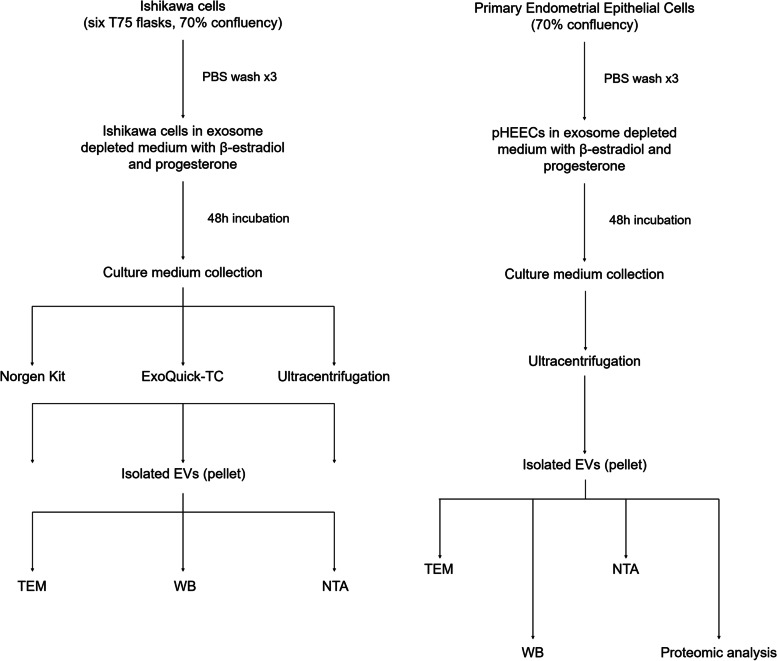
Methods: Ishikawa cells and pHEECs were in vitro cultured and hormonally treated; subsequently, conditioned medium was collected and EVs isolated. Ishikawa cells were used for the comparison of EVs isolation methods ultracentrifugation, ExoQuick-TC and Norgen Cell Culture Media Exosome Purification Kit (n = 3 replicates/isolation method). pHEECs were isolated from endometrial biopsies (n = 8/replicate; 3 replicates) collected from healthy oocyte donors with confirmed fertility, and protein content of EVs isolated by the most efficient methodology was analysed using liquid chromatography-tandem mass spectrometry. EV concentration and size were analyzed by nanoparticle tracking analysis, EV morphology visualized by transmission electron microscopy and protein marker expression was determined by Western blotting.
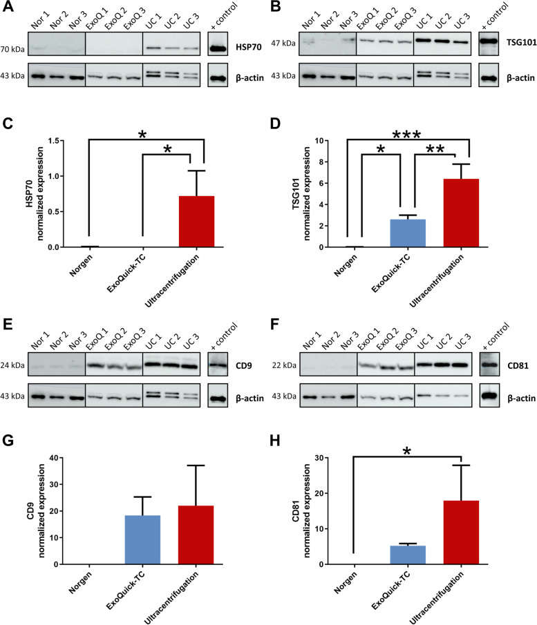
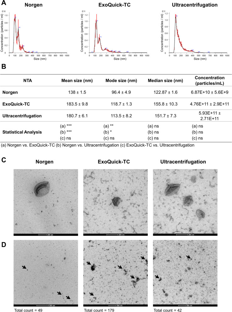
Results: Ultracentrifugation was the most efficient methodology for EV isolation from medium of endometrial epithelial cells. EVs secreted by pHEECs and isolated by ultracentrifugation were heterogeneous in size and expressed EV protein markers HSP70, TSG101, CD9, and CD81. Proteomic analysis identified 218 proteins contained in these EVs enriched in biological processes involved in embryo implantation, including cell adhesion, differentiation, communication, migration, extracellular matrix organization, vasculature development, and reproductive processes. From these proteins, 82 were selected based on their functional relevance in implantation success as possible implantation biomarkers.
Conclusions: EV protein cargos are implicated in biological processes related to endometrial receptivity, embryo implantation, and early embryo development, supporting the concept of a communication system between the embryo and the maternal endometrium via EVs. Identified proteins may define new biomarkers of endometrial receptivity and implantation success.

 求助内容:
求助内容: 应助结果提醒方式:


