Tulgar Toros, Murat Kayalar, Kemal Özaksar, Tahir Sadık Sügün, Yusuf Gürbüz
{"title":"Classification of vascularized fibular flap hypertrophy based on X-ray evaluation.","authors":"Tulgar Toros, Murat Kayalar, Kemal Özaksar, Tahir Sadık Sügün, Yusuf Gürbüz","doi":"10.5152/j.aott.2021.20206","DOIUrl":null,"url":null,"abstract":"<p><strong>Objective: </strong>The aim of this study was to analyze and classify hypertrophy seen in vascularized fibula flaps used for reconstruction of tubular bone defects.</p><p><strong>Methods: </strong>Thirty-three patients who underwent a vascularized fibula flap for the reconstruction of massive bone defects of the upper or lower extremity long bones were retrospectively reviewed and included in this study. There were 24 lower extremities (21 tibial and 3 femoral) and 9 upper extremities (4 humeral, 2 radial and 3 ulnar) reconstructions in this series. The mean age was 32.7 (range= 10- 59) years. The mean length of bony defect following initial debridement was 10.3 (range= 4-25) cm. The fibula was inserted as a single strut in 29 patients, and as a double barrel construct in 4 patients. The degree of fibular hypertrophy was calculated based on anteroposterior (AP) and lateral X-ray measurements of fibular flaps at an average postoperative period of 52 months. The difference in thickness between the initial and final x- ray measurements were expressed as percentage of hypertrophy. The variances seen in this period were defined and classified.</p><p><strong>Results: </strong>When bony consolidation of the 33 cases were examined in detail, 4 different modes of flap hypertrophy were defined: type 0- absence of hypertrophy, type 1- limited hypertrophy, type 2- marked hypertrophy triggered by stress fracture, and type 3- massive hypertrophy enhanced by peripheral bone production.</p><p><strong>Conclusion: </strong>Fibular hypertrophy follows different modes based on vascularity of the flap, amount of stress imparted on the flap, site of reconstruction, and whether the periosteal sleeve is retained at the reconstruction site. Determination of these factors at the initial period may help the surgeons to predict the final hypertrophy that will be seen at the end of flap maturation Level of Evidence: Level IV, Therapeutic Study.</p>","PeriodicalId":7097,"journal":{"name":"Acta orthopaedica et traumatologica turcica","volume":"55 6","pages":"541-546"},"PeriodicalIF":1.1000,"publicationDate":"2021-12-01","publicationTypes":"Journal Article","fieldsOfStudy":null,"isOpenAccess":false,"openAccessPdf":"https://www.ncbi.nlm.nih.gov/pmc/articles/PMC11583239/pdf/","citationCount":"0","resultStr":null,"platform":"Semanticscholar","paperid":null,"PeriodicalName":"Acta orthopaedica et traumatologica turcica","FirstCategoryId":"3","ListUrlMain":"https://doi.org/10.5152/j.aott.2021.20206","RegionNum":4,"RegionCategory":"医学","ArticlePicture":[],"TitleCN":null,"AbstractTextCN":null,"PMCID":null,"EPubDate":"","PubModel":"","JCR":"Q3","JCRName":"ORTHOPEDICS","Score":null,"Total":0}
引用次数: 0
Abstract
Objective: The aim of this study was to analyze and classify hypertrophy seen in vascularized fibula flaps used for reconstruction of tubular bone defects.
Methods: Thirty-three patients who underwent a vascularized fibula flap for the reconstruction of massive bone defects of the upper or lower extremity long bones were retrospectively reviewed and included in this study. There were 24 lower extremities (21 tibial and 3 femoral) and 9 upper extremities (4 humeral, 2 radial and 3 ulnar) reconstructions in this series. The mean age was 32.7 (range= 10- 59) years. The mean length of bony defect following initial debridement was 10.3 (range= 4-25) cm. The fibula was inserted as a single strut in 29 patients, and as a double barrel construct in 4 patients. The degree of fibular hypertrophy was calculated based on anteroposterior (AP) and lateral X-ray measurements of fibular flaps at an average postoperative period of 52 months. The difference in thickness between the initial and final x- ray measurements were expressed as percentage of hypertrophy. The variances seen in this period were defined and classified.
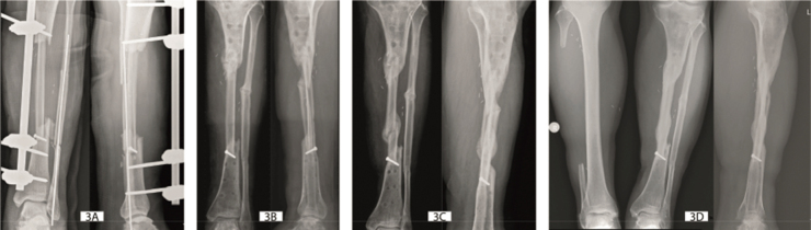
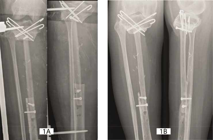
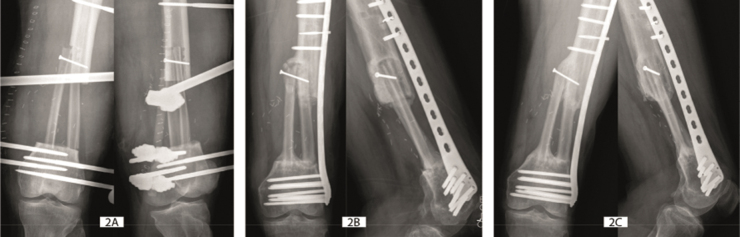
Results: When bony consolidation of the 33 cases were examined in detail, 4 different modes of flap hypertrophy were defined: type 0- absence of hypertrophy, type 1- limited hypertrophy, type 2- marked hypertrophy triggered by stress fracture, and type 3- massive hypertrophy enhanced by peripheral bone production.
Conclusion: Fibular hypertrophy follows different modes based on vascularity of the flap, amount of stress imparted on the flap, site of reconstruction, and whether the periosteal sleeve is retained at the reconstruction site. Determination of these factors at the initial period may help the surgeons to predict the final hypertrophy that will be seen at the end of flap maturation Level of Evidence: Level IV, Therapeutic Study.
期刊介绍:
Acta Orthopaedica et Traumatologica Turcica (AOTT) is an international, scientific, open access periodical published in accordance with independent, unbiased, and double-blinded peer-review principles. The journal is the official publication of the Turkish Association of Orthopaedics and Traumatology, and Turkish Society of Orthopaedics and Traumatology. It is published bimonthly in January, March, May, July, September, and November. The publication language of the journal is English.
The aim of the journal is to publish original studies of the highest scientific and clinical value in orthopedics, traumatology, and related disciplines. The scope of the journal includes but not limited to diagnostic, treatment, and prevention methods related to orthopedics and traumatology. Acta Orthopaedica et Traumatologica Turcica publishes clinical and basic research articles, case reports, personal clinical and technical notes, systematic reviews and meta-analyses and letters to the Editor. Proceedings of scientific meetings are also considered for publication.
The target audience of the journal includes healthcare professionals, physicians, and researchers who are interested or working in orthopedics and traumatology field, and related disciplines.
 求助内容:
求助内容: 应助结果提醒方式:


