Dunya Moradi, Reza Eyvazpour, Fariborz Rahimi, Ali Jahan, Seyed Hossein Rasta, Mahdad Esmaeili
{"title":"Electroencephalographic Activity in Patients with Claustrophobia: A Pilot Study.","authors":"Dunya Moradi, Reza Eyvazpour, Fariborz Rahimi, Ali Jahan, Seyed Hossein Rasta, Mahdad Esmaeili","doi":"10.4103/jmss.JMSS_62_20","DOIUrl":null,"url":null,"abstract":"<p><strong>Background: </strong>Exposure to small confined spaces evokes physiological responses such as increased heart rate in claustrophobic patients. However, little is known about electrocortical activity while these people are functionally exposed to such phobic situations. The aim of this study was to examine possible changes in electrocortical activity in this population.</p><p><strong>Method: </strong>Two highly affected patients with claustrophobia and two healthy controls participated in this <i>in vivo</i> study during which electroencephalographic (EEG) activity was continuously recorded. Relative power spectral density (rPSD) was compared between two situations of being relaxed in a well-lit open area, and sitting in a relaxed chair in a small (90 cm × 180 cm × 155 cm) chamber with a dim light. This comparison of rPSDs in five frequency bands of EEG was intended to investigate possible patterns of change in electrical activity during fear-related situation. This possible change was also compared between claustrophobic patients and healthy controls in all cortical areas.</p><p><strong>Results: </strong>Statistical models showed that there is a significant interaction between groups of participants and experimental situations in all frequency bands (<i>P</i> < 0.01). In other words, claustrophobic patients showed significantly different changes in electrical activity while going from rest to the test situation. Clear differences were observed in alpha and theta bands. In the theta band, while healthy controls showed an increase in rPSD, claustrophobic patients showed an opposite decrease in the power of electrical activity when entering the confined chamber. In alpha band, both groups showed an increase in rPSD, though this increase was significantly higher for claustrophobic patients.</p><p><strong>Conclusion: </strong>The effect of <i>in vivo</i> exposure to confined environments on EEG activity is different in claustrophobic patients than in healthy controls. Most of this contrast is observed in central and parietal areas of the cortex, and in the alpha and theta bands.</p>","PeriodicalId":37680,"journal":{"name":"Journal of Medical Signals & Sensors","volume":"11 4","pages":"262-268"},"PeriodicalIF":1.1000,"publicationDate":"2021-10-20","publicationTypes":"Journal Article","fieldsOfStudy":null,"isOpenAccess":false,"openAccessPdf":"https://ftp.ncbi.nlm.nih.gov/pub/pmc/oa_pdf/39/48/JMSS-11-262.PMC8588885.pdf","citationCount":"2","resultStr":null,"platform":"Semanticscholar","paperid":null,"PeriodicalName":"Journal of Medical Signals & Sensors","FirstCategoryId":"1085","ListUrlMain":"https://doi.org/10.4103/jmss.JMSS_62_20","RegionNum":0,"RegionCategory":null,"ArticlePicture":[],"TitleCN":null,"AbstractTextCN":null,"PMCID":null,"EPubDate":"2021/10/1 0:00:00","PubModel":"eCollection","JCR":"Q4","JCRName":"ENGINEERING, BIOMEDICAL","Score":null,"Total":0}
引用次数: 2
Abstract
Background: Exposure to small confined spaces evokes physiological responses such as increased heart rate in claustrophobic patients. However, little is known about electrocortical activity while these people are functionally exposed to such phobic situations. The aim of this study was to examine possible changes in electrocortical activity in this population.
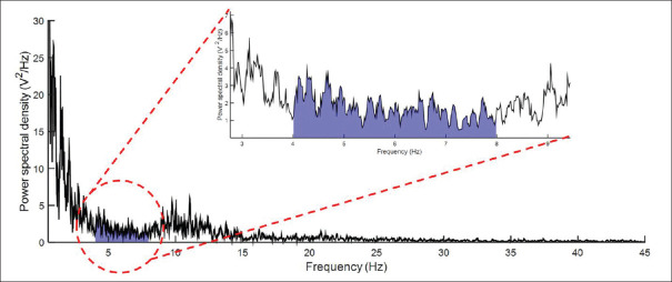
Method: Two highly affected patients with claustrophobia and two healthy controls participated in this in vivo study during which electroencephalographic (EEG) activity was continuously recorded. Relative power spectral density (rPSD) was compared between two situations of being relaxed in a well-lit open area, and sitting in a relaxed chair in a small (90 cm × 180 cm × 155 cm) chamber with a dim light. This comparison of rPSDs in five frequency bands of EEG was intended to investigate possible patterns of change in electrical activity during fear-related situation. This possible change was also compared between claustrophobic patients and healthy controls in all cortical areas.
Results: Statistical models showed that there is a significant interaction between groups of participants and experimental situations in all frequency bands (P < 0.01). In other words, claustrophobic patients showed significantly different changes in electrical activity while going from rest to the test situation. Clear differences were observed in alpha and theta bands. In the theta band, while healthy controls showed an increase in rPSD, claustrophobic patients showed an opposite decrease in the power of electrical activity when entering the confined chamber. In alpha band, both groups showed an increase in rPSD, though this increase was significantly higher for claustrophobic patients.
Conclusion: The effect of in vivo exposure to confined environments on EEG activity is different in claustrophobic patients than in healthy controls. Most of this contrast is observed in central and parietal areas of the cortex, and in the alpha and theta bands.
背景:暴露于狭小的密闭空间会引起生理反应,如幽闭恐惧症患者心率加快。然而,当这些人功能性地暴露在这种恐惧情境中时,对皮层电活动知之甚少。本研究的目的是检查这一人群中皮层电活动的可能变化。方法:对2例重度幽闭恐惧症患者和2例健康对照者进行体内实验,连续记录脑电图(EEG)活动。相对功率谱密度(rPSD)比较了在光线充足的开放区域放松和在光线昏暗的小房间(90 cm × 180 cm × 155 cm)中坐在放松的椅子上的两种情况。通过对脑电图5个频带rpsd的比较,探讨恐惧相关情境下脑电活动变化的可能模式。我们还比较了幽闭恐惧症患者和健康对照者在所有皮质区域的这种可能的变化。结果:统计模型显示,各频段被试组与实验情境之间存在显著交互作用(P < 0.01)。换句话说,幽闭恐惧症患者在从休息状态到测试状态的过程中,脑电活动表现出明显不同的变化。在α和θ波段观察到明显的差异。在theta波段,健康对照组的rPSD增加,而幽闭恐惧症患者在进入密闭的房间时,电活动的功率却相反地下降。在α波段,两组的rPSD均有所增加,但幽闭恐惧症患者的rPSD明显增加。结论:密闭环境对幽闭恐惧症患者脑电图活动的影响与健康对照不同。这种对比主要发生在皮层的中央和顶叶区域,以及α和θ波段。
期刊介绍:
JMSS is an interdisciplinary journal that incorporates all aspects of the biomedical engineering including bioelectrics, bioinformatics, medical physics, health technology assessment, etc. Subject areas covered by the journal include: - Bioelectric: Bioinstruments Biosensors Modeling Biomedical signal processing Medical image analysis and processing Medical imaging devices Control of biological systems Neuromuscular systems Cognitive sciences Telemedicine Robotic Medical ultrasonography Bioelectromagnetics Electrophysiology Cell tracking - Bioinformatics and medical informatics: Analysis of biological data Data mining Stochastic modeling Computational genomics Artificial intelligence & fuzzy Applications Medical softwares Bioalgorithms Electronic health - Biophysics and medical physics: Computed tomography Radiation therapy Laser therapy - Education in biomedical engineering - Health technology assessment - Standard in biomedical engineering.


 求助内容:
求助内容: 应助结果提醒方式:


